Methods for detecting abnormal epithelial tissue

a technology of epithelial tissue and abnormality, applied in the field of abnormal epithelial tissue detection, can solve the problems of significant relative death hazards of patients who delay a cancer consultation for at least two months, and the delay is not only awkward, but also may be impossibl

Inactive Publication Date: 2006-10-26
ZILA PHARMA
View PDF14 Cites 3 Cited by
  • Summary
  • Abstract
  • Description
  • Claims
  • Application Information

AI Technical Summary

Problems solved by technology

Patients who delay in obtaining a cancer consultation for at least two months have significantly higher relative hazards of death than do patients with a shorter delay.
The selective visualization of abnormal mucosal tissue using such light sources is hindered by normal ambient light (daylight or normal artificial light) falling upon the tissue being examined, such that the standard procedure for conducting such examinations calls for darkening the room in which the examination is conducted.
This is not only awkward but also may be impossible when the examination is conducted in rooms with large window areas or when other procedures on other patients are being conducted in the same room served by common conventional lighting.

Method used


Image

  • Methods for detecting abnormal epithelial tissue
  • Methods for detecting abnormal epithelial tissue
  • Methods for detecting abnormal epithelial tissue

Examples


example 1

[0009] A routine visual examination of the oral cavity is made, noting the presence of any lesions on the attached gingiva, the buccal mucosa, the floor of the mouth, the hard and soft palate and the dorsal, lateral and ventral tongue. Any lesions noted by this routine examination are recorded.

example 2

[0010] After completing the routine examination of Example 1, the patient is then instructed to rinse the mouth with a 1% acetic acid solution for up to one minute and then expectorate.

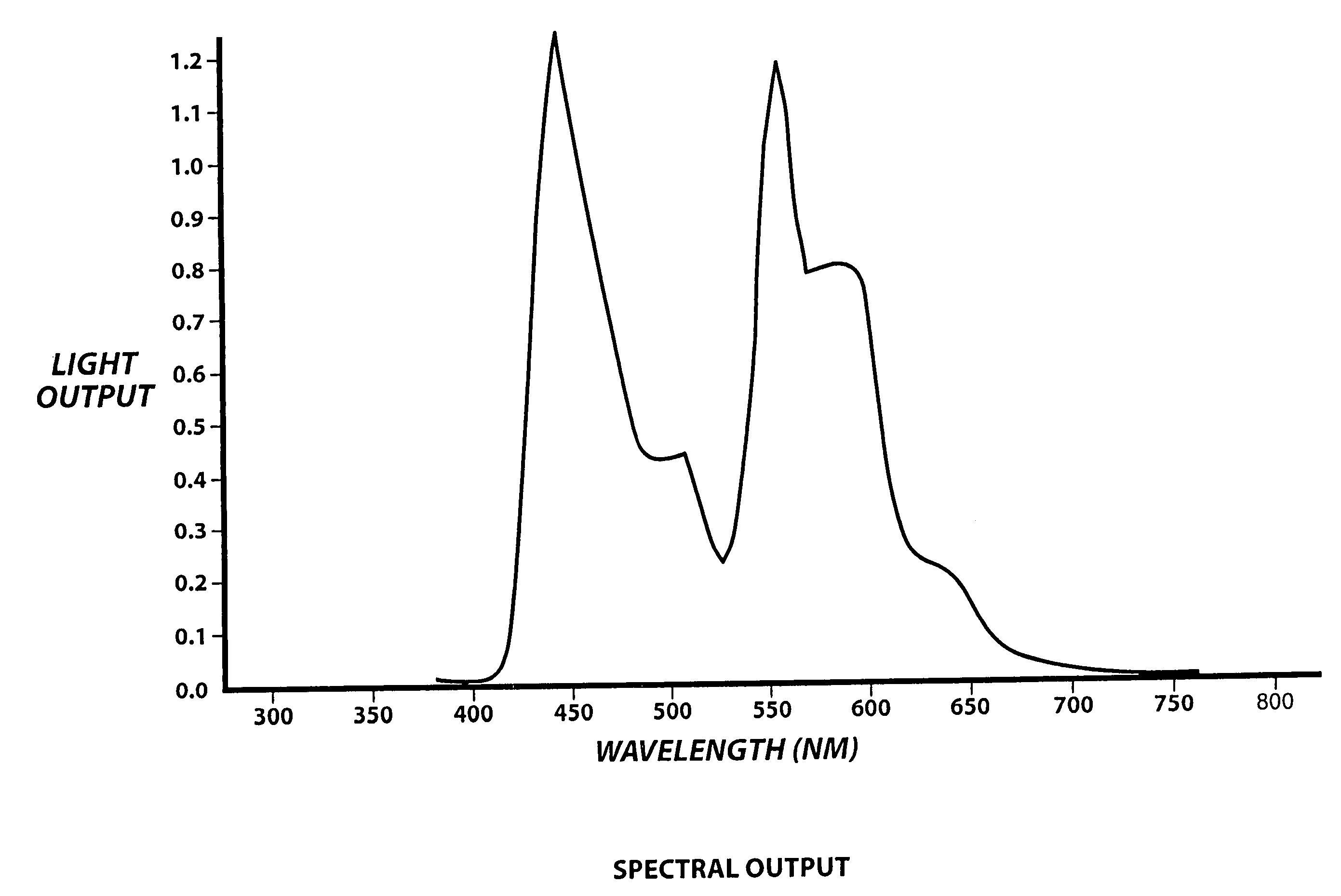
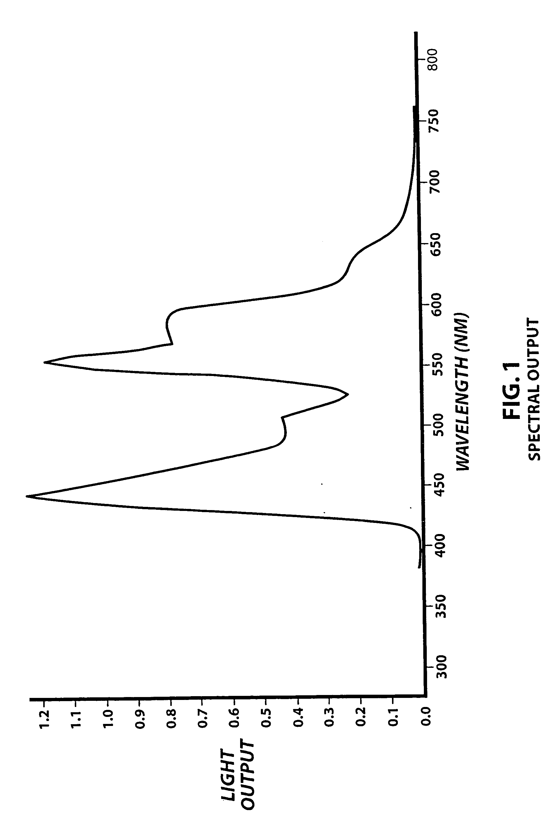
[0011] The chemiluminescent light source described in the Lonky patent U.S. Pat. No. 5,329,938, commercially available under the registered trademark VIZILITE®, is activated by bending the flexible outer capsule, breaking the brittle inner vial. The capsule is then shaken and it is inserted into the retractor. The light provided has spectral peaks at about 450 nm, 550 nm and a smaller peak in the red region at about 600 nm, as indicated in FIG. 1. These spectral peaks produce a bluish-white light.

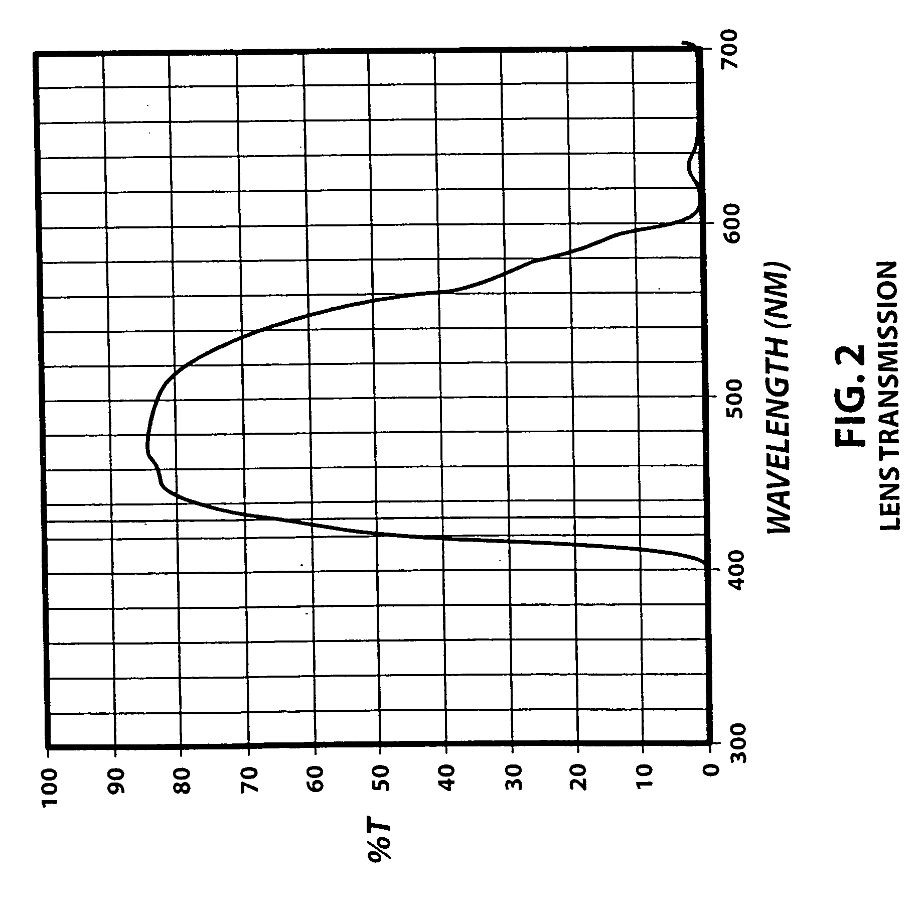
[0012] The examining clinician then dons a pair of spectacles provided with lens which only transmit light in the wavelength band of 400-600 nm, as indicated in FIG. 2. These spectacles are shaped to minimize illumination reaching the examiner's eyes from above and from the sides. These spectacles are avail...


PUM


Abstract

The visibility of abnormal tissue under light having wavelength peaks which selectively identify abnormal tissue is enhanced in the presence of normal ambient light by viewing the tissue through lens which transmit the wavelength peaks but block transmission of other wavelengths.

Description

[0001] This invention relates to methods for detecting abnormal epithelial tissue, which may harbor tumor phenotypes. [0002] In another respect the invention pertains to improved methods for conducting real time in vivo examinations of epithelial tissue to detect abnormalities which may be cancerous or which may eventually develop invasive cancer. BACKGROUND OF THE INVENTION [0003] Patients who delay in obtaining a cancer consultation for at least two months have significantly higher relative hazards of death than do patients with a shorter delay. Thus, if patients are more regularly subjected to effective cancer screening, the mortality risks of cancer would be reduced. Thus, there was a need for a simple, rapid screening test for detecting abnormal mucosal tissue which may harbor tumor phenotypes, which may indicate the presence of or the eventual development of invasive cancer. [0004] Abnormal epithelial tissue can be visually identified and located in real time in vivo using sel...

Claims


Application Information

Patent Timeline
IPC IPC(8): A61B6/00
CPCA61B5/0059A61B5/445A61B5/444A61B5/0088
Inventor BRIDE, MARK
Owner ZILA PHARMA
Why Eureka
Try Eureka