
Automatic image contrast in computer aided diagnosis

a technology of automatic image contrast and computer aided diagnosis, applied in image enhancement, image analysis, instruments, etc., can solve the problems of poor image quality, adversely affecting the percentage of correct results obtained from the cad system, and improper positioning of patients

Pending Publication Date: 2006-05-25
CARESTREAM HEALTH INC
View PDF10 Cites 19 Cited by
  • Summary
  • Abstract
  • Description
  • Claims
  • Application Information

AI Technical Summary

Problems solved by technology

A number of factors can adversely influence the percentage of correct results obtained from the CAD system.
Errors can result from factors such as poor image quality, improper positioning of the patient, film variations, scanner performance, obscuration from fibroglandular tissue, and other problems.
Because of these difficulties, some view the success rate in correctly identifying and diagnosing microcalcification structures as disappointing.
The percentage of false negative (FN) and false positive (FP) errors is still too high when using conventional CAD systems.
However, even using advanced neural networks and other powerful image analysis and decision-making tools may only provide incremental improvement over existing methods.
One problem of particular interest for accurate diagnosis and optimized image display is the need for a suitable adjustment in image contrast.
Thus, even where adjustments are “easy to use” from a GUI perspective, there can be disadvantages and problems when these features are not used well.
While some systems have employed adjustment utilities and techniques for the adjustment task, there is still considerable dissatisfaction with the adjustment task and with its outcome.
Some practitioners dislike the job of manually adjusting image contrast and find the various contrast adjustment tools confusing, using only a portion of the interface as a result.
Providing more complex, capable tools may improve potential accuracy but may not be well accepted by medical professionals, particularly those trained on earlier equipment where contrast, from one image to the next, had been relatively constant for images from the same imaging system.
One problem with applying conventional image contrast adjustment techniques to mammography images is that specific details needed by the diagnostician may be lost.
This complicates the task of contrast adjustment, whether done manually by the imaging system operator or performed automatically for an image.

Method used


Image

  • Automatic image contrast in computer aided diagnosis
  • Automatic image contrast in computer aided diagnosis
  • Automatic image contrast in computer aided diagnosis

Examples


Embodiment Construction

[0046] The present description is directed in particular to elements forming part of, or cooperating more directly with, apparatus in accordance with the invention. It is to be understood that elements not specifically shown or described may take various forms well known to those skilled in the art.

[0047] The method of the present invention can use hardware and software components, but is independent of any particular component characteristics such as architecture, operating system, or programming language, for example. In general, the type of system equipment that is conventionally employed for scanning, processing, and classification of mammography image data, or of other types of medical image data, is well known and includes at least some type of computer or computer workstation, having a logic processor which may be dedicated solely to the assessment and maintenance of medical images or may be used for other data processing functions in addition to image processing. Typically,...


PUM


Abstract

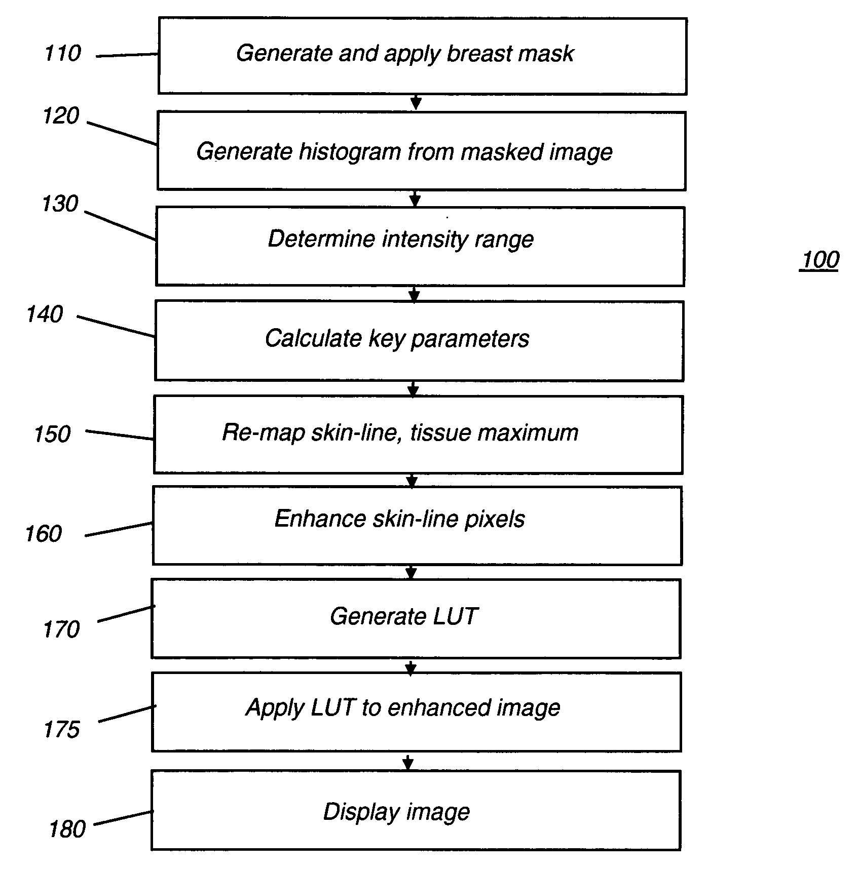
A method for adjusting contrast level for displaying an image, particularly for computed aided diagnosis. The method includes the steps of: masking one or more portions of the image to obtain a cropped image; forming a histogram of pixel intensity values from the cropped image using a plurality of bins, each bin having a predetermined bin width; designating one or more bins as image background bins and mapping pixels in the image background bins to a display background value; designating bins for upper and lower bound values and mapping tissue bins, having values between upper and lower bound values, to tissue display values, forming a contrast-adjusted image thereby; assigning pixels along the contour of the cropped image to one or more skin line bins; mapping pixels in the one or more skin line bins to an enhanced pixel value in the contrast-adjusted image; and displaying the contrast-adjusted image.

Description

RELATED APPLICATIONS [0001] Reference is made to, and priority is claimed from, U.S. Provisional Application No. 60 / 631,156, entitled “AUTOMATIC IMAGE CONTRAST IN CAD APPLICATION”, filed on Nov. 24, 2004 in the names of Zheng et al, and which is assigned to the assignee of this application, and incorporated herein by reference.FIELD OF THE INVENTION [0002] The present invention generally relates to medical image analysis and more particularly relates to an automated method for setting contrast level for display and analysis of a medical image. BACKGROUND OF THE INVENTION [0003] The benefits of computer-aided diagnosis in radiology in general, and particularly in mammography, are widely recognized. There has been efforts directed toward computer-aided methods that assist the diagnostician to correctly and efficiently identify problem areas detected in a mammography image and to improve the accuracy with which diagnoses are made using this information. [0004] In mammography, it is rec...

Claims


Application Information

Patent Timeline
Patent Type & Authority Applications(United States)
IPC IPC(8): G06K9/00
CPCG06T5/009G06T5/40G06T2207/10116G06T2207/20012G06T2207/20132G06T2207/20148G06T2207/20192G06T2207/30068G06T2207/30096G06T7/136G06T5/92
Inventor ZHANG, DAOXIAN H.ZHENG, YANG
Owner CARESTREAM HEALTH INC
Learn More