
Frozen tissue microarray

a tissue microarray and frozen technology, applied in the field of tissues analysis, can solve the problems of difficult to create tissue microarray cores with a consistent depth in the recipient block, paraffin-processed tissue cores, and may not be the best option to demonstrate specific antigens,

Inactive Publication Date: 2005-12-22
COVANCE
View PDF4 Cites 16 Cited by
  • Summary
  • Abstract
  • Description
  • Claims
  • Application Information

AI Technical Summary

Benefits of technology

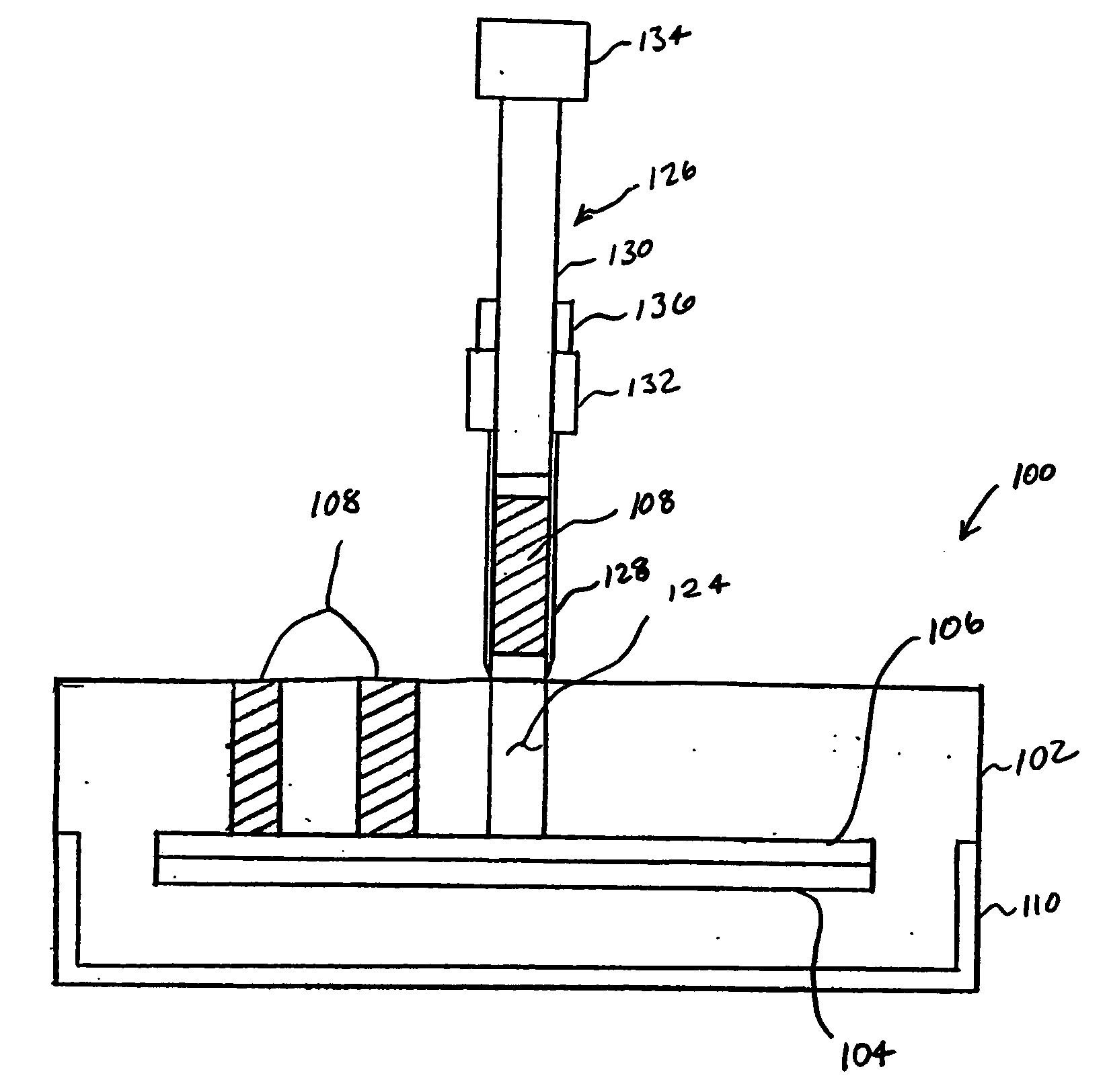
[0010] This and other needs are met by the present invention, which in accord with one aspect includes a tissue microarray having frozen tissue cores that extend from the top of the tissue microarray to a release. The release is a material from which embedding material of the tissue microarray can easily be removed. A stiffener is used with the release to maintain a flat shape of the release. The tissue cores may be formed from whole (i.e., paraffin-free) frozen tissue. By using the release, cores of frozen embedding material can be easily removed from the tissue microarray during the process of inserting donor tissue cores into the embedding material of the tissue microarray.
[0014] In still another aspect of the present, invention, a moisture barrier is provided for protecting the micrometer and slide mechanisms of a microarrayer from moisture and frost damage during production of tissue microarrays.

Problems solved by technology

A problem associated with the creation of tissue microarrays using the apparatus and method described above is the removal of frozen embedding material from the recipient block using the recipient punch.
Another problem associated with the creation of tissue microarrays is the difficulty in creating cores in the recipient block that have a consistent depth.
Paraffin-processed tissue cores, however, may not be the best option to demonstrate specific antigens.
However, the techniques used to control the size of the cores have problems when used with whole frozen tissues as the ability of an user to detect when a core fully inserted into a recipient block is compromised when the core is formed from whole frozen tissue.
When thawing of the frozen material occurs, the user inserting the core into the recipient block does not detect exactly when the bottom of the core contacts the bottom of the hole in the recipient block because the thawed material flexes.
Since the user cannot detect when the core is fully inserted, the core can be over-inserted into the hole in the recipient block, which results in the core being crushed.

Method used


Image

  • Frozen tissue microarray
  • Frozen tissue microarray
  • Frozen tissue microarray

Examples


Embodiment Construction

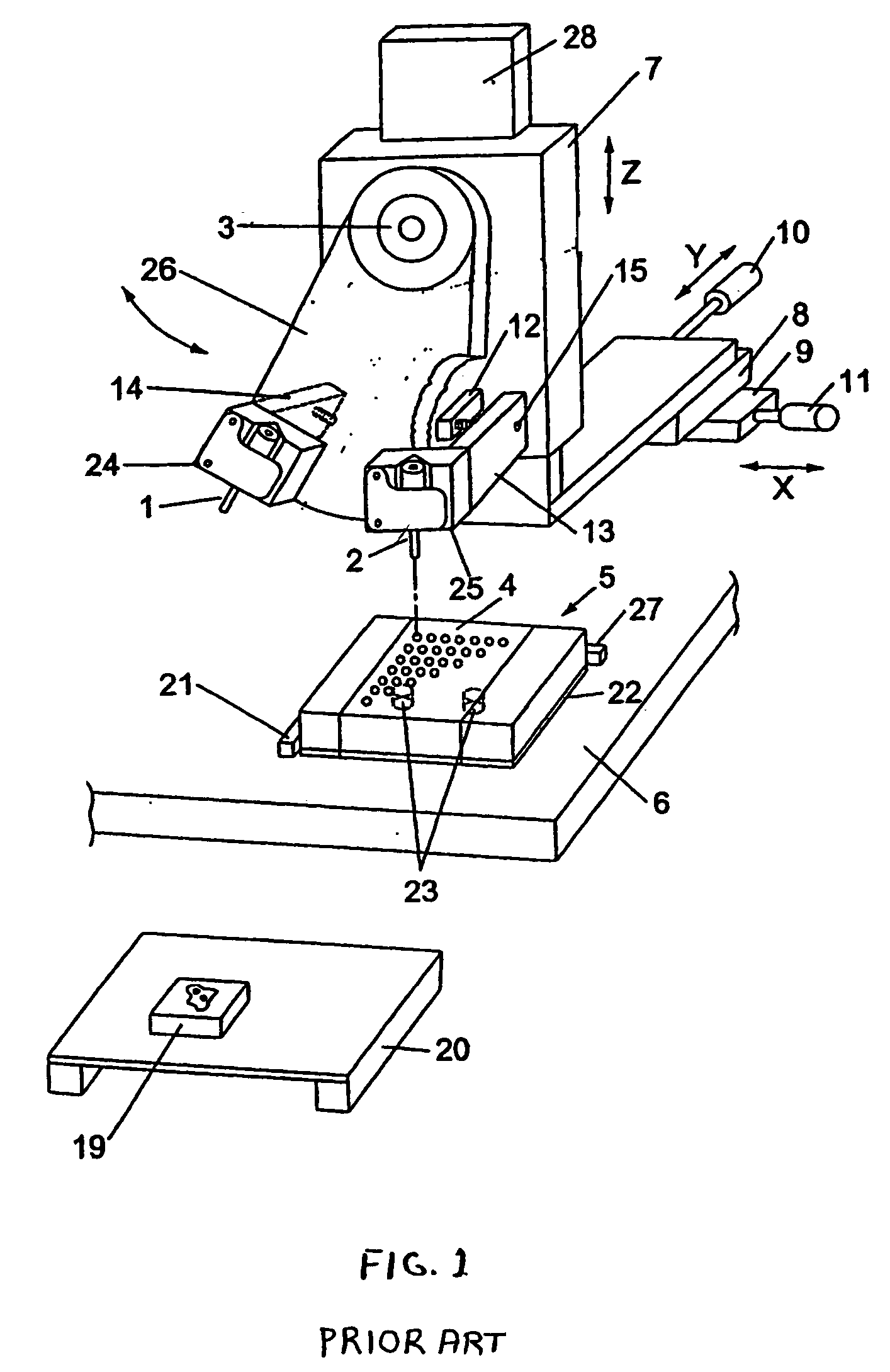
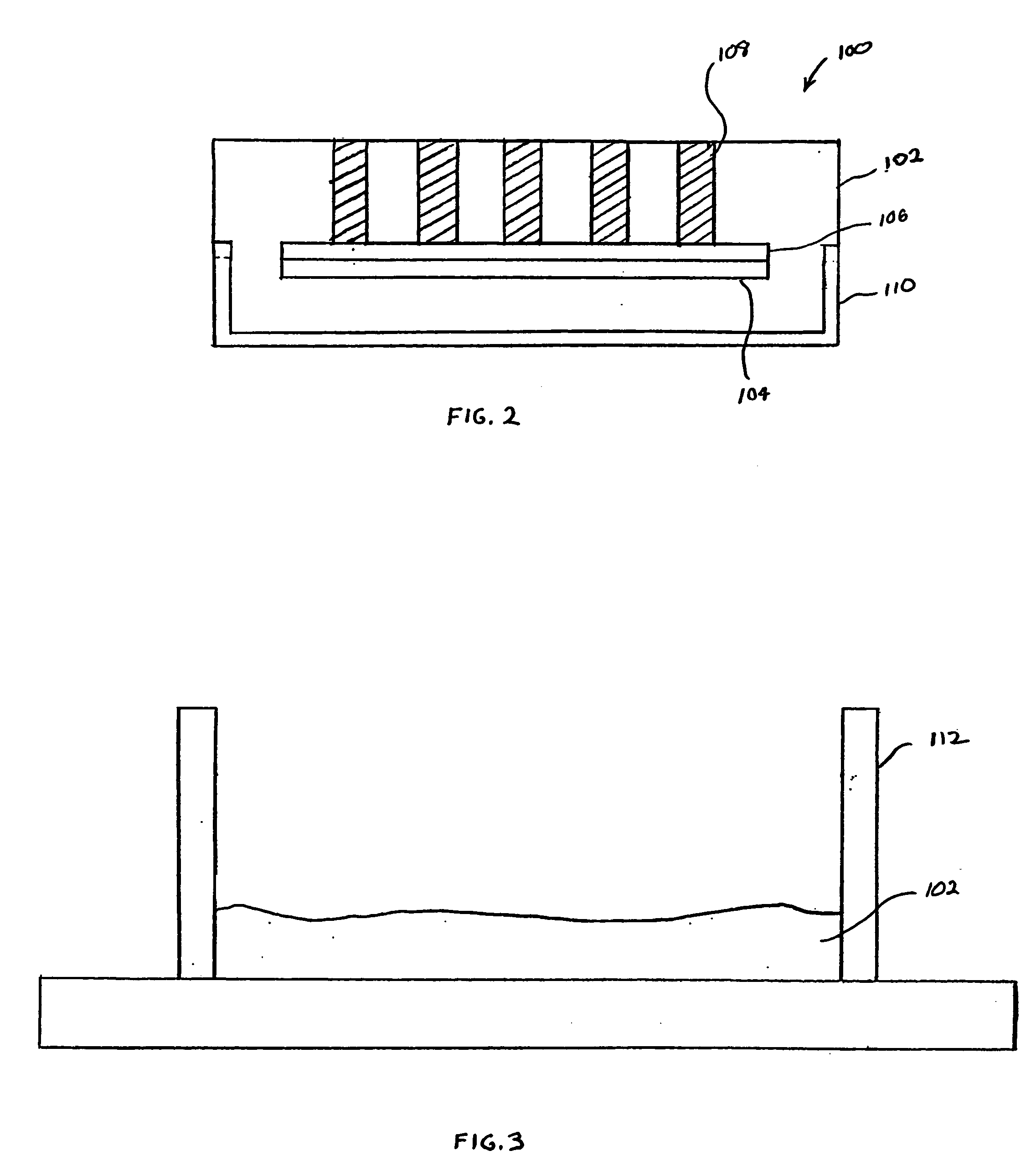
[0028]FIG. 2 illustrates a tissue microarray 100 according to an aspect of the invention. Consistent with conventional tissue microarrays, the tissue microarray 100 includes embedding material 102 positioned with a cassette 110, and the tissue microarray 100 is not limited as to a particular type of cassette 110. The cassette 110 is a fixture that is held by a tissue microarrayer (shown in FIG. 1) when tissue cores 108 are being inserted into the embedding material 102 of the tissue microarray 100. Many types of embedding material 102 exist, and the tissue microarray is not limited as to a particular embedding material 102. For example, the embedding material 102, also known as optimal cutting temperature compound (OCT), can formed from such materials as paraffin wax, nitrocellulose, polyvinyl alcohol, carbowax (polyethylene glycols), gelatin and agar.

[0029] The tissue microarray 100 also includes a release 106 and a stiffener 104. The release 106 and stiffener 104 may be a single ...


PUM

PropertyMeasurementUnit
diameteraaaaaaaaaa
diameteraaaaaaaaaa
diameteraaaaaaaaaa

Abstract

A tissue microarray includes frozen tissue cores that extend from the top of the tissue microarray to a release. The release is a material from which embedding material of the tissue microarray can easily be removed. A stiffener is used with the release to maintain a flat shape of the release. The tissue cores may be formed from whole (i.e., paraffin-free) frozen tissue. A method and tool for manufacturing the tissue microarray are also disclosed.

Description

FIELD OF THE INVENTION [0001] The invention relates generally to analysis of tissues and, more specifically, to an improved method and apparatus for forming frozen tissue microarrays. BACKGROUND OF THE INVENTION [0002] Tissue microarrays increase the throughput of molecular analyses by simultaneously arraying proteins, nucleic acids, and other biomolecules. The tissue microarrays allow for the study of protein expression, antigen localization and molecular profiling of tissues samples. These tissue microarrays typically include cylindrical biopsies of human or animal tissue taken from already fixed paraffin-embedded tissue blocks or fresh frozen tissues. The cylindrical biopsies are the positioned into a tissue recipient microarray block, which can then be histologically prepared to produce sections for subsequent study. Tissue microarrays can provide a great number of tissues to be screened on a single slide and allow for rapid, large scale assessment of tissues compared to convent...

Claims


Application Information

Patent Timeline
Patent Type & Authority Applications(United States)
IPC IPC(8): C12M1/00C12M1/16C12M1/18G01N1/36G01N1/42G01N33/48
CPCB01J2219/00286B01J2219/00313B01J2219/00315B01J2219/0074B01L3/5085G01N2001/368B01L2300/0819G01N1/08G01N1/36G01N1/42B01L2200/12
Inventor POSTOYALKO, ANDREWROBERTS, JOHN
Owner COVANCE
Learn More