
System, method and medium for acquiring and generating standardized operator independent ultrasound images of fetal, neonatal and adult organs

Active Publication Date: 2005-11-10
EASTERN VIRGINIA MEDICAL SCHOOL
View PDF22 Cites 71 Cited by
  • Summary
  • Abstract
  • Description
  • Claims
  • Application Information

AI Technical Summary

Benefits of technology

[0012] It is a feature and advantage of the present invention to provide a system, method and medium that can be used to acquire and generate standardized operator independent ultrasound images of fetal, neonatal and / or adult organs.
[0013] It is still another feature and advantage of the present invention to provide a system, method and medium that can be used to acquire and generate standardized operator independent ultrasound images of standard anatomic planes of fetal, neonatal and / or adult organs that can be used to detect normal and / or abnormal imaging relationships within the organ.
[0014] It is yet another feature and advantage of the present invention to provide a system, method and medium that can be used to improve the efficiency and diagnostic capabilities of current ultrasound examinations of fetal, neonatal and / or adult organs.
[0015] It is a further feature of the present invention to facilitate sonography-related teaching and education, and facilitate training of various medical personnel.
[0016] At least one embodiment of the present invention can utilize, for example, a computer program in conjunction with, for example, a general purpose computer and / or standard sonography equipment to obtain and optionally display 2D, 3D and / or 4D ultrasound images. In addition, at least one embodiment of the present invention can provide a medical evaluation or diagnosis of aspects of fetal, neonatal and adult organs (e.g., the fetal heart).
[0017] In an exemplary method in accordance with the present invention, a reference plane is obtained for a particular body organ, which can be used as a baseline from which to obtain other planes of interest, such as the four-chamber view plane of the fetal heart. The reference plane can optionally be a standard representative plane that is relatively easy to obtain on 2D ultrasonography, such as the four-chamber view plane of the fetal heart. Exemplary reference planes for the fetal head are the axial biparietal diameter, the axial posterior fossa, the axial lateral ventricles, and the coronal corpus callosum.

Problems solved by technology

However, even for trained personnel, 3D volume manipulation by the multiplanar display process can be difficult to perform, particularly when the volume involves relatively complex anatomical organs, such as the central nervous system or the heart.
Furthermore, in obstetrical ultrasound imaging, the variable position of the fetus within the uterus is an added factor that raises the level of difficulty.
This manual process of generating images in obstetrical sonography results in a lack of standardization, image recognition and consistency of diagnosis.
Thus, despite the advantages that volume sonography may provide, significant limitations still exist in volume sonography today.
The acquisition, display and manipulation of 3D volumes require a substantial learning curve.
Volume sonography in obstetrics is still hampered by the lack of uniformity due to the variable position of the fetus within the uterus and by the technical skills needed to retrieve diagnostic 2D planes out of a 3D volume.
These difficulties are compounded for 3D volumes involving anatomically complex organs such as the fetal brain and / or heart.
The lack of standardization in the acquisition and display of 3D volumes is an impediment to training.

Method used


Image

  • System, method and medium for acquiring and generating standardized operator independent ultrasound images of fetal, neonatal and adult organs
  • System, method and medium for acquiring and generating standardized operator independent ultrasound images of fetal, neonatal and adult organs
  • System, method and medium for acquiring and generating standardized operator independent ultrasound images of fetal, neonatal and adult organs

Examples


Embodiment Construction

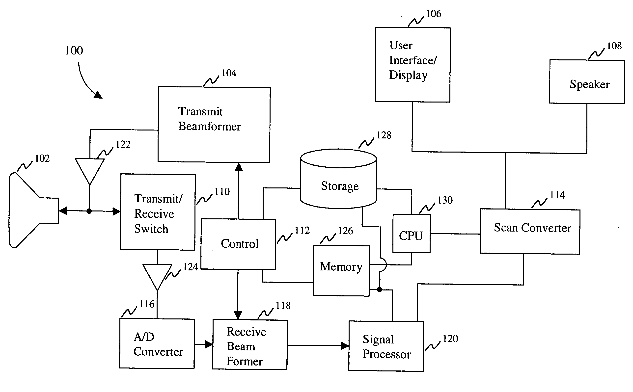
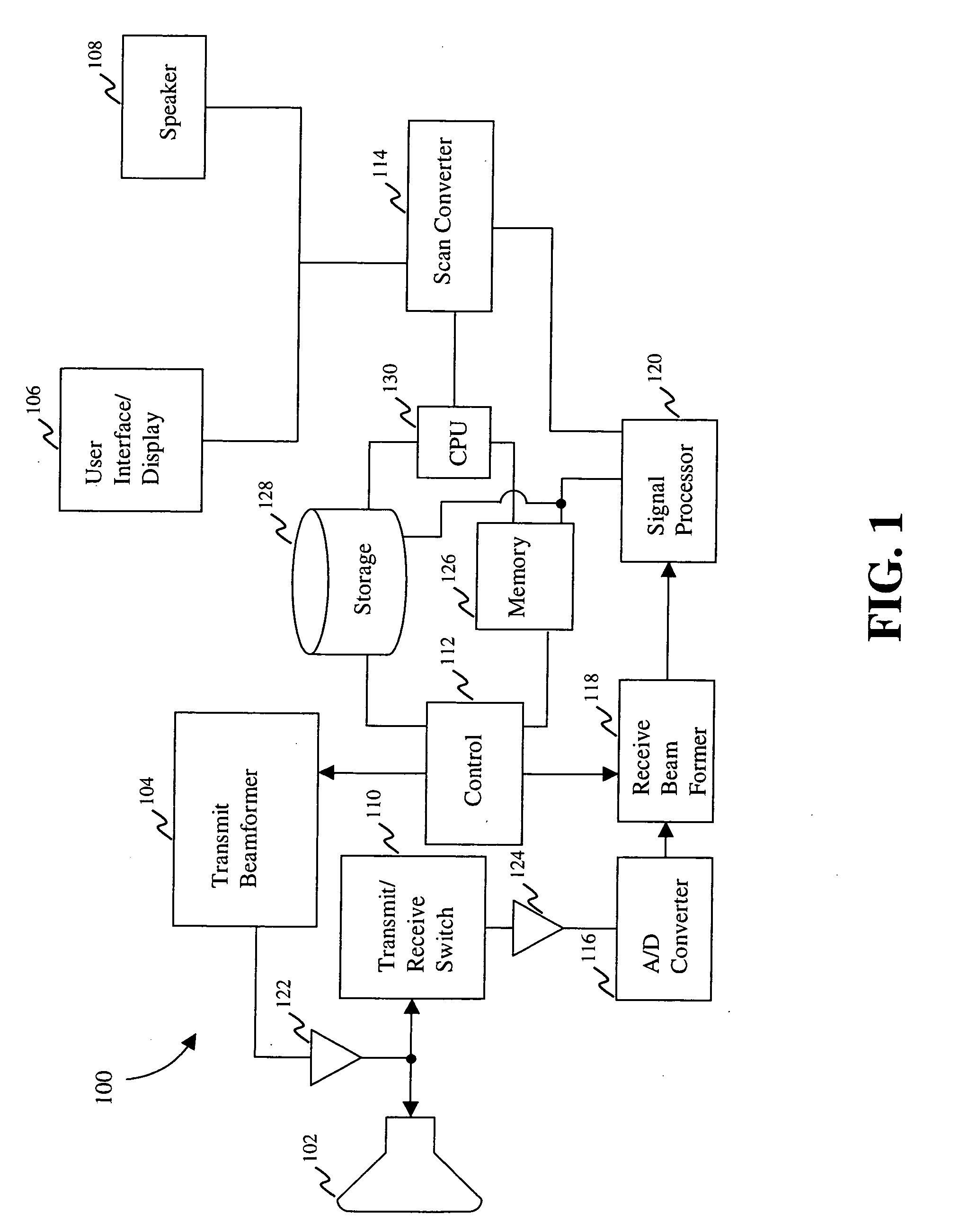
[0073] Before explaining at least one embodiment of the invention in detail, it is to be understood that the invention is not limited in its application to the details of construction and to the arrangements of the components set forth in the following description or illustrated in the drawings. The invention is capable of other embodiments and of being practiced and carried out in various ways. Also, it is to be understood that the phraseology and terminology employed herein are for the purpose of description and should not be regarded as limiting.

[0074] As such, those skilled in the art will appreciate that the conception, upon which this disclosure is based, may readily be utilized as a basis for the designing of other structures, methods and systems for carrying out the several purposes of the present invention. It is important, therefore, that the invention be regarded as including equivalent constructions to those described herein insofar as they do not depart from the spirit...


PUM


Abstract

A system, method and medium for standardizing a manner of acquisition and display of ultrasound images. In one embodiment of the invention, a volumetric image of an organ is acquired in a standardized manner. Relationships such as formulas are utilized to automatically generate anatomical planes of interest within the volume that can be displayed independent of the user.

Description

CROSS-REFERENCE TO RELATED APPLICATIONS [0001] This application claims priority to, and incorporates by reference, Provisional Application Ser. No. 60 / 463,045, filed Apr. 16, 2003. This application is a continuation-in-part application of Ser. No. 10 / 823,793, filed Apr. 14, 2004.DESCRIPTION BACKGROUND OF THE INVENTION FIELD OF THE INVENTION [0002] The present invention generally relates to acquiring and generating ultrasound still and / or real time images and, more particularly, to acquiring and generating operator-independent displays of ultrasound still and / or real time images of standard anatomic planes of fetal, neonatal and / or adult organs. BACKGROUND DESCRIPTION [0003] In the art of medical imaging, the development of three-dimensional (3D) and four-dimensional (4D) ultrasonography provides an advance in imaging technology. With 3D ultrasonography, an infinite number of two-dimensional (2D) planes are acquired of (or within) a target volume. The volume acquired by 3D ultrasonog...

Claims


Application Information

Patent Timeline
IPC IPC(8): A61B8/14
CPCA61B8/08A61B8/0866A61B8/0883A61B8/0891G06T2210/41A61B8/463A61B8/523G06T7/0012G06T2207/30048A61B8/14
Inventor ABUHAMAD, ALFRED Z.
Owner EASTERN VIRGINIA MEDICAL SCHOOL
Learn More