Topographic coatings and coating methods for medical devices

a technology of topographic coatings and medical devices, applied in the direction of colloidal chemistry, prosthesis, blood vessels, etc., can solve the problems of destroying or removing any therapeutic agent deposited thereon, and achieve the effect of facilitating the manipulation of medical devices

Inactive Publication Date: 2005-10-13
XTENT INC
View PDF99 Cites 179 Cited by
  • Summary
  • Abstract
  • Description
  • Claims
  • Application Information

AI Technical Summary

Benefits of technology

[0004] The present invention provides implantable medical devices coated with topographic coatings and methods for the manufacture and use thereof. Such topographic coatings are useful for various purposes. First, such coatings may be configured to provide channels, apertures, holes, depressions, reservoirs and other suitable structures to contain therapeutic agents. In addition, such topographic coatings may be configured to protect those regions of the medical device on which therapeutic agents are deposited to prevent damage or removal of therapeutic agents due to contact during assembly, handling, or delivery to a treatment site. Further, such topographic coatings may be used to facilitate manipulation of the medical device by a delivery instrument or catheter.
[0006] One or more therapeutic agents, including anti-restenosis, anti-proliferative, immunosuppressive, antibiotic, thrombolytic, cytotoxic, cystostatic, and other agents, as well as growth factors, DNA, and other substances, may be deposited in the regions of low elevation in the topographic coating. These agents may be deposited with only a solvent which evaporates off, or may be mixed with a durable or bioerodable carrier to provide a delivery matrix for the agent. Different agents and / or different concentrations of the same agent may be deposited in different regions at various locations on the medical device, or within the same region in vertical layers or side-by-side deposits. Further, the topographic coating itself may be mixed, infused or impregnated with a therapeutic agent the same or different than that deposited in the regions of low elevation. Additional layers of polymers, metals, ceramics, proteins, or other materials may be applied to the medical device either over or under the topographic coating and / or therapeutic agent. Such layers may be used to protect the underlying material from damage or removal, to control elution rates of therapeutic agents in the underlying material, to promote adhesion of overlying material to the underlying surface, and other purposes. Such therapeutic agents and other materials may be deposited by spraying, syringe coating, dipping, vacuum deposition, sputtering, and other methods, but preferably such agents and materials are deposited using jet printing, which allows for highly precise deposition in a predetermined pattern coordinated with the pattern of the high and low elevation regions in the topographic coating.

Problems solved by technology

Because these stent valves may contact the outer surface of the stent segments, they have the potential to damage or remove any therapeutic agent deposited thereon.

Method used


Image

  • Topographic coatings and coating methods for medical devices
  • Topographic coatings and coating methods for medical devices
  • Topographic coatings and coating methods for medical devices

Examples


second embodiment

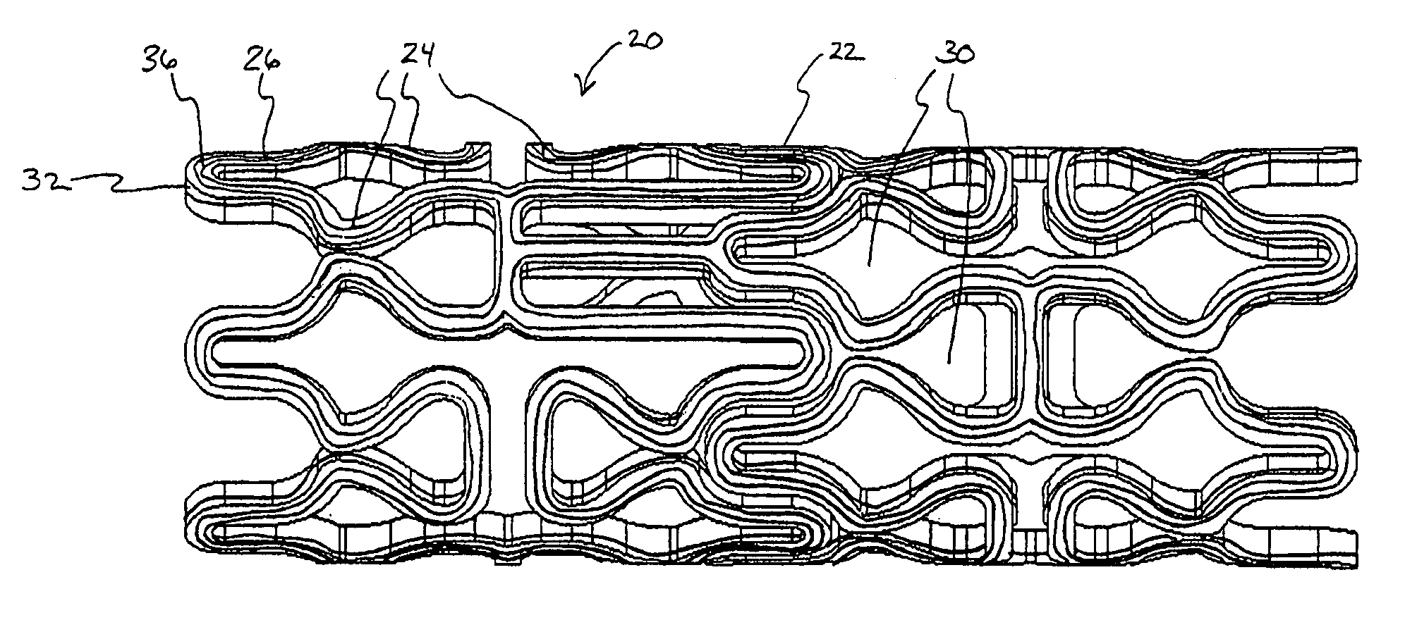
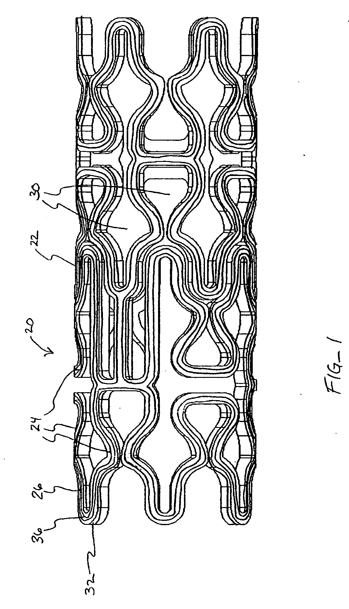
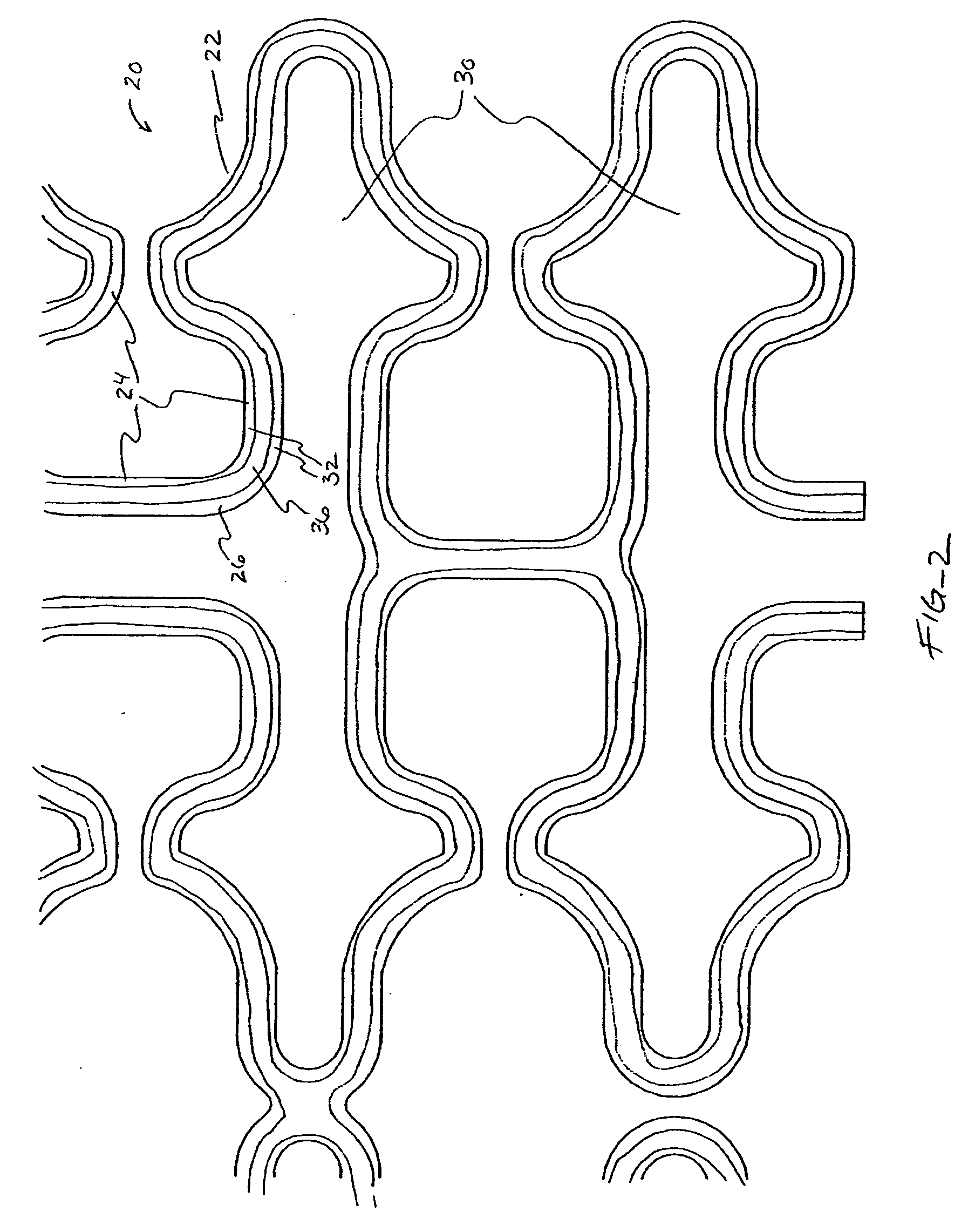
[0041] In a second embodiment, shown in FIGS. 4A-B, topographic layer 32 is formed in a single ridge 38 on outer surface 26 generally parallel to the struts 24, forming two low elevation regions 40 on either side of ridge 38. A therapeutic agent may then be deposited in either or both low elevation regions 40.

third embodiment

[0042] In a third embodiment, shown in FIGS. 5A-5B, topographic layer 32 is formed in a single ridge 42 covering substantially all of outer surface 26. Topographic layer 32 may be impregnated or mixed with a therapeutic agent that elutes from it at a desired rate. Alternatively, reservoirs, depressions, concavities, holes or other regions of low elevation may be formed in ridge 42 by masking during deposition or after deposition by drilling, heating, cutting, etching, or other methods, as illustrated in FIGS. 8A-B below. A therapeutic agent may then be deposited in the low elevation regions. As a further alternative, topographic layer 32 may be used to facilitate manipulation of the stent in a stent delivery catheter, as described more fully below.

[0043] Referring now to FIGS. 6-7, in a further embodiment, topographic layer 32 is formed in a plurality of bumps 44 in a predetermined pattern on outer surface 26. In this embodiment, topographic layer 32 may be comprised of a therapeuti...


PUM


Abstract

Medical devices having topographic coatings are provided. The topographic coatings have regions of high and low elevation and may be composed of polymers, metals, ceramics, proteins and other biocompatible materials. Such topographic coatings facilitate the deposition, elution, and protection of therapeutic agents on the medical device, manipulation of the medical device, and other purposes. In particularly preferred embodiments, the medical device comprises a stent for vascular implantation.

Description

CROSS-REFERENCES TO RELATED APPLICATIONS [0001] The present application is a non-provisional of U.S. Patent Application Ser. No. 60 / 561,041 (Attorney Docket No. 021629-002600), filed Apr. 9, 2004, the full disclosure of which is incorporated herein by reference.BACKGROUND OF THE INVENTION [0002] Coronary stents are tubular scaffolds deployed in stenotic lesions in diseased coronary arteries to maintain the patency of the arterial lumen. Uncoated (or bare metal) coronary stents have suffered from a significant incidence of restenosis, the recurrence of stenotic plaque in the lesion where a stent has been placed. In recent years, coronary stents coated with therapeutic agents such as paclitaxel, rapamycin, or various analogs thereof have shown success in preventing restenosis. In such drug-eluting stents, the therapeutic agent is typically mixed with a durable or bioerodable polymer and applied to the stent by dipping, spraying, or syringe dispensing. However, such techniques suffer f...

Claims


Application Information

Patent Timeline
IPC IPC(8): A61F2/00A61F2/82
CPCA61F2/91A61F2/915A61F2002/828A61F2002/91508A61F2002/91516A61F2230/0013A61F2002/91533A61F2002/9155A61F2002/91591A61F2250/0068A61F2210/0076A61F2002/91525
Inventor GRAINGER, JEFFRY J.HNOJEWYJ, OLEXANDER
Owner XTENT INC
Why Eureka
Try Eureka