
Particle agglutination detection method and device

a particle agglutination and detection method technology, applied in the field of receptorligand interaction detection, can solve the problems of not teaching or suggesting art, and achieve the effect of simple use, fast and accurate results, and avoiding false positive results

Inactive Publication Date: 2005-04-21
INVERNESS MEDICAL SWITZERLAND GMBH
View PDF18 Cites 8 Cited by
  • Summary
  • Abstract
  • Description
  • Claims
  • Application Information

AI Technical Summary

Benefits of technology

[0081] An additional further advantage of the present invention is that it is optionally and preferably suitable for both blood grouping and for cross-matching.
[0082] A feature of the present invention is that the direction of flow of the test components is preferably perpendicular to the surface of a porous body.
[0083] An advantage of the present invention is that the method and device are simple to use.
[0084] A further advantage of the present invention is that false positive results are avoided.
[0085] A further advantage of the present invention is that results are obtained quickly and accurately, preferably with a very low margin of error.
[0086] In the present specification, a number of terms are discussed hereinunder, for the purposes of description only and without any intention of being limiting. Unless otherwise defined, all technical and scientific terms used herein have the same meaning as commonly understood by one of ordinary skill in the art to which this invention belongs.

Problems solved by technology

The background art does not teach or suggest a quick, simple, and accurate blood testing procedure involving an easily portable device, based on perpendicular movement through filters of defined pore sizes, use of which requires little or no specialized training, and which provides directly recordable results.

Method used


Image

  • Particle agglutination detection method and device
  • Particle agglutination detection method and device

Examples


example 1

Wash Solutions

[0143] Wash solutions used in generating the examples were:

[0144] Dulbecco's Phosphate Buffered Saline (PBS), obtained from Biological Industries, Beit Ha'emek, Israel.

[0145] A solution made from 1:1 diluted PBS in water with 4% w / v Poly Ethylene Glycol (PEG) 15000-20000MW (Fluka) and 0.3% w / v dextran sulfate sodium salt (Amersham Biosciences).

[0146] A. Dulbecco's Phosphate Buffered Saline (PBS) with 0.001-0.01% w / v polyoxyethylene-10-tridecyl ether (Sigma).

example 2

Blood Grouping

[0147] A 0.5×0.5 cm piece of Ahlstrom #142 filter paper was placed on an absorbent pad. Two μL of whole blood were pipetted in the center of the filter and followed by 2 μL of an anti-blood group reagent (Gamma Biologicals Inc., Hosuton, Tex., USA). The filter was then washed with a few drops (from a dropper bottle) of a wash solution and dried. Alternatively, a 4 mm diameter circle of the Ahlstrom #142 filter paper, 10 μL of blood and 10 μL of the anti-blood group reagent were used followed by few drops of wash solution C from Example 1.

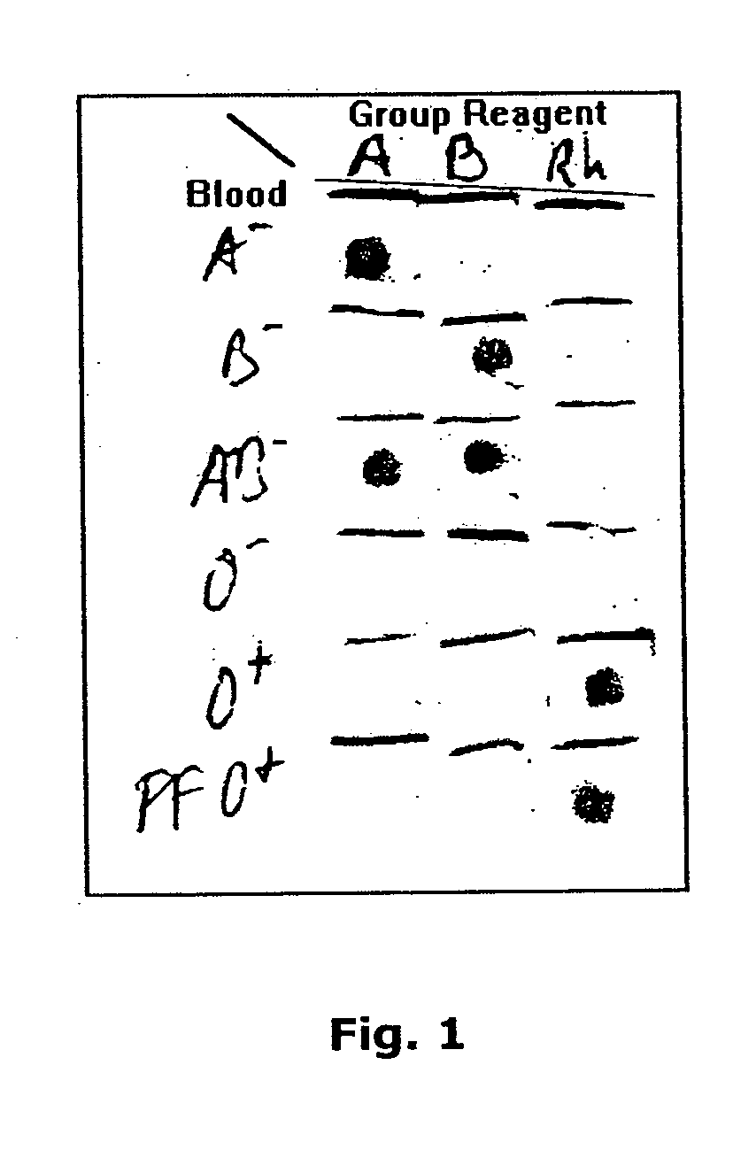
[0148] This test was repeated with multiple blood specimens of various blood groups (received from and pre-tested by the Central Blood Services, Israel Red Magen David, Tel Hashomer, Israel) and each of these was tested with anti-A, anti-B, anti-AB and anti-D (=Rh) blood group reagents. The A− blood generated a red spot only with anti-A and anti-AB reagents. The B− blood generated a spot with the anti-B and anti-AB reagents. The AB− ...

example 3

Blood Grouping with Dried Reagents

[0149] 0.5×0.5 cm pieces of Ahlstrom #142 filter paper were placed on a non absorbent surface (bottom of empty, disposable Petri dishes). A 50 μL aliquot of an anti-blood group reagent (Gamma Biologicals Inc., Hosuton, Tex., USA) was pipetted on each of the pieces of filter paper. The Petri dishes with the anti-blood group reagents containing filter pieces were incubated overnight at 37° C. The filter pieces were dry at that time.

[0150] For testing the ability of the dried, antibody impregnated pieces to correctly identify the blood grouping of whole blood specimens, they were placed on an absorbent pad and a 1 μL aliquot of the test blood was placed in the approximate center of each of a series of filter pads. The series included pads, each impregnated with either one of anti-A, anti-B or anti-D (=Rh) reagent.

[0151] Excess blood was washed from the filter pads by placing 5-10 drops of either wash solution A or B. The filter pieces were then air ...


PUM

PropertyMeasurementUnit
Flow rateaaaaaaaaaa
Concentrationaaaaaaaaaa
Volumeaaaaaaaaaa

Abstract

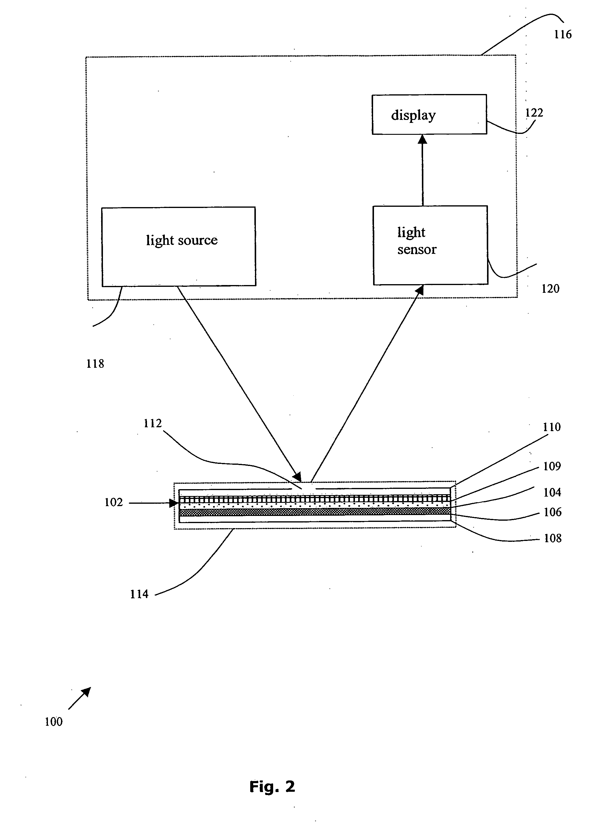
A method for the detection and / or visualization of particle agglutination, comprising: placing a volume of a suspension of the particles on a filter, the filter being constructed so as to permit passage of individual particles; placing a volume of a solution or suspension containing an agglutinating agent at the location of the particle suspension; optionally placing a wash solution at the location of the agglutinating agent; and observing the surface at the location for the presence of particles, such presence indicating that agglutination of the particles occurred. There is also provided a method for detection of agglutination reactions in general and hemagglutination reactions, such as used in blood grouping and cross-matching, in particular. The method is comprised of successive vertical additions of whole blood, blood grouping reagent and wash to a filter. In case of hemagglutination a colored, preferably red or reddish dot becomes visible after washing. A device and a kit based on the invention is also claimed and facilitates blood grouping and matching in non-laboratory environment without the need for laboratory instruments.

Description

[0001] This application claims the benefit of priority from U.S. Provisional Patent Application No. 60 / 485,118, filed Jul. 8, 2003.FIELD OF THE INVENTION [0002] The present invention relates to detection of receptor-ligand interactions in general, and more particularly, to the detection of blood group antigens and their antibodies for the purpose of blood typing and matching as employed in transfusion medicine. BACKGROUND OF THE INVENTION [0003] Particle agglutination is a widely adopted immunological method for the detection and visualization of antigen-antibody interaction due to its simplicity, rapidity and relative sensitivity (Riochet 1993). Cells in general, and red blood cells in particular, are particles which are amenable to a variety of agglutination methods. As such, agglutination of red blood cells (hemagglutination) is employed for the detection of antigens on their surface and antibodies to such antigens in blood group typing or matching as significant components in pr...

Claims


Application Information

Patent Timeline
IPC IPC(8): C12Q1/68G01N33/543G01N33/80
CPCG01N33/80
Inventor ROTT, GENNADYSAMUELS, FREDFISH, FALK
Owner INVERNESS MEDICAL SWITZERLAND GMBH
Learn More