
Method and system for spectral examination of vascular walls through blood during cardiac motion

a vascular wall and spectral examination technology, applied in the field of spectral examination of vascular walls through blood during cardiac motion, can solve the problems of difficult to isolate the spectral response of the vessel wall or other structures of interest, the probe head is poorly controlled, and the treatment and/or analysis of the vessel wall through blood are complicated further, so as to improve the treatment effect of the vessel wall and improve the effect of the treatmen

Active Publication Date: 2005-02-24
INFRAREDX INC
View PDF12 Cites 27 Cited by
  • Summary
  • Abstract
  • Description
  • Claims
  • Application Information

AI Technical Summary

Benefits of technology

[0009] One approach to addressing the influence of motion is electrocardiographic gating (ECG gating), where some pre-selected segment of the cardiac cycle is used for taking measurements. ECG gating can enable spectral acquisition during a period of lesser movement, or may reduce the variance in the distance between the vessel and the catheter head between samples by collecting information at the some specified point in the cycle over multiple cardiac cycles. ECG gating, however, has had limited success due to the weak correlation between vessel motion and catheter motion.
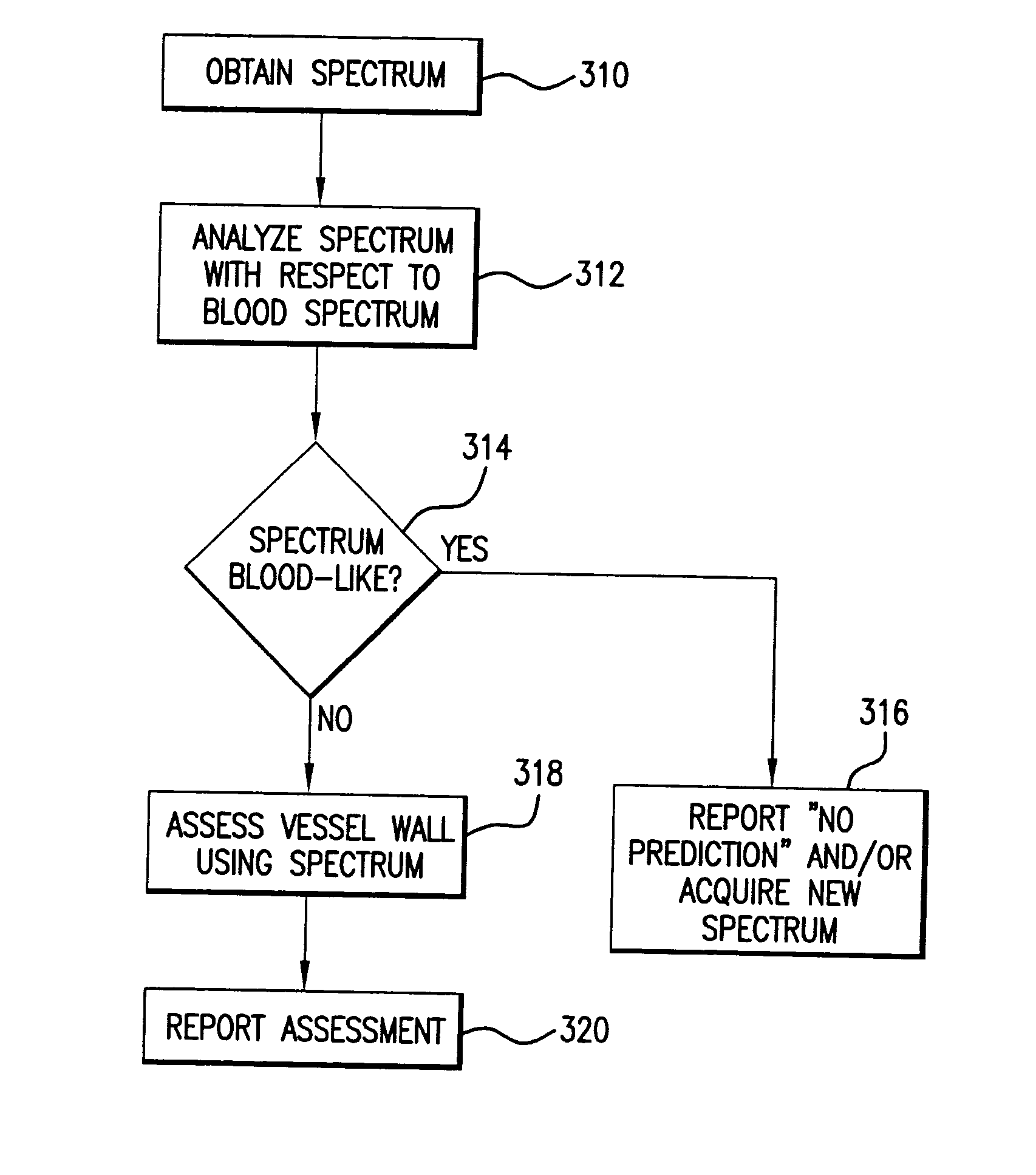
[0011] The present invention concerns an approach for improving the treatment and / or examination of vessels walls through fluid, such as blood. In the specific example, the invention is used for near infrared (NIR) spectroscopy. The invention can take advantage of the probe, such as catheter head, motion by identifying the points in time when the head is closest to the vessel wall or farthest from the vessel wall. Identification of this relative location enables meaningful spectral readings in larger vessels. In short, instead of trying to overcome motion (e.g., by centering the catheter), this approach takes advantage of motion by identify times when the catheter is closer to the vessel wall, in order to gather more useful spectral information or improve the efficacy of the treatment of the vessel walls.

Problems solved by technology

The treatment and / or analysis of vessel walls through blood, however, are complicated further by the fact that the physical relationship between the probe, and typically the probe head, and the blood vessel walls is poorly controlled.
Thus, in diagnostic applications, such as spectroscopy applications, the unknown pathlength will affect the degree to which the detected spectrum is dominated by unwanted signal sources, such as blood, thus making it difficult to isolate the spectral response of the vessel walls or other structures of interest.
The problem is further complicated in large vessels since the catheter head can be so far from the vessel walls that it is impossible to acquire any useful information.
ECG gating, however, has had limited success due to the weak correlation between vessel motion and catheter motion.
These approaches are clinically undesirable, however.
Among other problems, this may cause all signals to be poor in a large vessel, due to the large catheter-to-vessel distance, however.

Method used


Image

  • Method and system for spectral examination of vascular walls through blood during cardiac motion
  • Method and system for spectral examination of vascular walls through blood during cardiac motion
  • Method and system for spectral examination of vascular walls through blood during cardiac motion

Examples


first embodiment

[0095]FIG. 5 shows the catheter providing for increased movement during operation.

[0096] The catheter head 58 includes a high density mass or weight 118 that is located away from the axis of rotation 116 for the catheter head 58. As a result, when the catheter 56 is driven to rotate, see arrow 110, the catheter head 58 traces an elliptical path 112. This elliptical path 112 brings the optical window 48 of the catheter head 58 into close proximity with the target 22. By timing the capture of the optical signal and assessment of the vessel walls with the point in time when the elliptical path 112 of the catheter head 58 brings the optical window 48 in proximity to the target area 22, the signal to noise is maximized.

[0097] As discussed previously, this rotation is sometimes combined with other simultaneous operations, as described previously, in which catheter head 58 is initially placed at the distal point of the vessel portion to be evaluated, and then pulled back through the vesse...

second embodiment

[0098]FIGS. 6A and 6B show the catheter providing for increased movement during operation.

[0099] Referring to 6A, here the catheter head 58 includes right and left wings or fins 114A and 114B. These wings or fins 114A, 114B interact with the flowing blood 108 to cause the catheter head to move relative to the target area 22. This causes the optical window 48 to periodically move closer to the target area 22 to enable high signal to noise analysis of the target area 22 by reducing the pathlength through the blood 108.

[0100] In the specific embodiment, the wings 114A, 114B are angled relative to the centerline of the catheter head 58 as shown in FIG. 6B. Here the flowing blood 108 acts on the wings 114 to push the catheter head 58 in a direction that is orthogonal to the optical window 48, in the direction of the target area 22. Thus, as the flow rate of the blood 108 increases, the catheter head is moved closer to the target area 22.

[0101] Generally, the wings may be designed accor...


PUM


Abstract

A method for improving the treatment and / or examination of vessel walls through fluid, such as blood, functions by identifying the points in time when the catheter is closest to the vessel wall or farthest from the vessel wall. Identification of this relative location enables improved spectral readings in larger vessels. In short, instead of trying to overcome motion (e.g., by centering the catheter), this approach takes advantage of motion by identify times when the catheter is closer to the vessel wall, in order to gather more useful spectral information or improve the efficacy of the treatment of the vessel walls. In the specific example, the invention is used for near infrared (NIR) spectroscopy. In some embodiments, the catheter head is designed to induce relative movement between the head and the vessel walls.

Description

BACKGROUND OF THE INVENTION [0001] Probe-based, such as catheter-based, optical systems are applicable to a number of diagnostic and therapeutic medical applications. Optical coherence tomography is used to provide spatial resolution, enabling the imaging of internal structures. Spectroscopy is used to characterize the composition of structures, enabling the diagnosis of medical conditions, by differentiating between cancerous, dysplastic, and normal tissue structures, for example. Ablation systems are used to remove or destroy structures within the body to address various diseases, such as tachycardias, tumors, and coronary artery disease, in another example of a probe-based optical system. [0002] For example, in one specific spectroscopic application, an optical source, such as a tunable laser, is used to access or scan a spectral band of interest, such as a scan band in the near infrared or 750 nanometers (nm) to 2.5 micrometers (μm). The generated light is used to illuminate tis...

Claims


Application Information

Patent Timeline
Patent Type & Authority Applications(United States)
IPC IPC(8): A61B5/00
CPCA61B5/0075A61B5/7285A61B5/0086A61B5/0084
Inventor CAPLAN, JAY D.MARSHIK-GEURTS, BARBARA J.
Owner INFRAREDX INC
Learn More