
Single catheter system that provides both intravascular ultrasound and fluorescence lifetime imaging

a single catheter and lifetime imaging technology, applied in the field of catheter-based vascular imaging systems, can solve the problems of limited imaging penetration depth, inability to reliably determine the size of deep plaque cores or outward remodeling, and inability to detect rupture-prone thin-cap fibroatheromas (tcfas),

Active Publication Date: 2022-06-07
RGT UNIV OF CALIFORNIA
View PDF5 Cites 0 Cited by
  • Summary
  • Abstract
  • Description
  • Claims
  • Application Information

AI Technical Summary

Benefits of technology

Enables precise characterization of atherosclerotic vessels by combining high-resolution IVUS and FLIm data, improving plaque vulnerability assessment without the need for exogenous contrast agents and facilitating standard clinical use.

Problems solved by technology

Unfortunately, the above-described intravascular imaging techniques have a number of shortcomings.
IVUS is not able to detect rupture-prone thin-cap fibroatheromas (TCFAs) due to limited spatial resolution (>100 μm).
Conversely, OCT has a spatial resolution of 10-20 μm and can detect thin caps and can also assess inflammation, but has limited imaging penetration depth and cannot reliably determine the size of deep plaque cores or outward remodeling, which are important indicators of plaque vulnerability.
NIRS has the ability to detect lipid cores, but has limited sensitivity for other proteins of interest, such as collagen and elastin.
However, the only previously existing intravascular catheter is a research tool, which is only able to gather FLIm and IVUS images sequentially.
This makes it hard to co-register the FLIm and IVUS images because during heartbeats, the catheter translates longitudinally within the blood vessel.
These registrations issues can limit the effectiveness of combining FLIm and IVUS images to identify vulnerable plaques within a blood vessel.

Method used


Image

  • Single catheter system that provides both intravascular ultrasound and fluorescence lifetime imaging
  • Single catheter system that provides both intravascular ultrasound and fluorescence lifetime imaging
  • Single catheter system that provides both intravascular ultrasound and fluorescence lifetime imaging

Examples


Embodiment Construction

[0024]The following description is presented to enable any person skilled in the art to make and use the present embodiments, and is provided in the context of a particular application and its requirements. Various modifications to the disclosed embodiments will be readily apparent to those skilled in the art, and the general principles defined herein may be applied to other embodiments and applications without departing from the spirit and scope of the present embodiments. Thus, the present embodiments are not limited to the embodiments shown, but are to be accorded the widest scope consistent with the principles and features disclosed herein.

[0025]The data structures and code described in this detailed description are typically stored on a computer-readable storage medium, which may be any device or medium that can store code and / or data for use by a computer system. The computer-readable storage medium includes, but is not limited to, volatile memory, non-volatile memory, magneti...


PUM

PropertyMeasurementUnit
sizeaaaaaaaaaa
optical fluorescenceaaaaaaaaaa
lengthsaaaaaaaaaa

Abstract

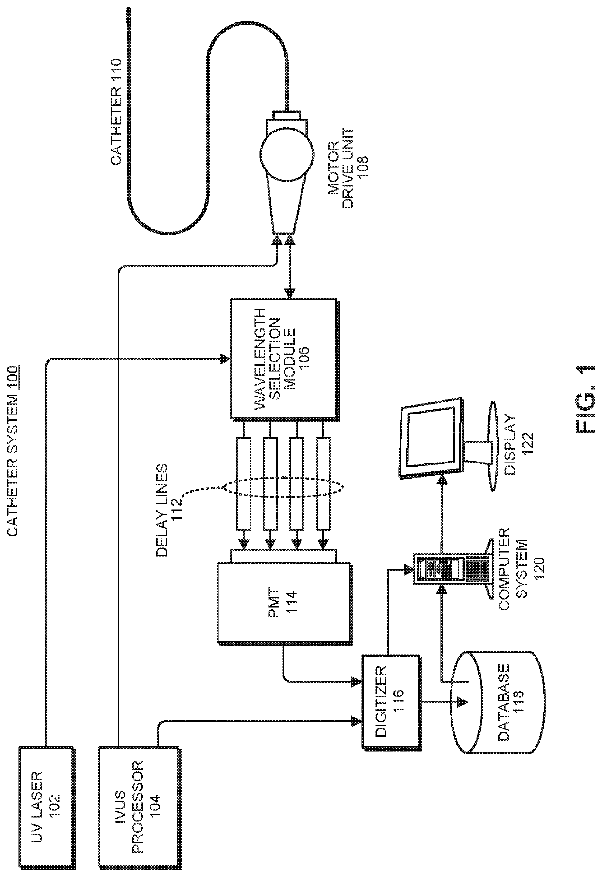
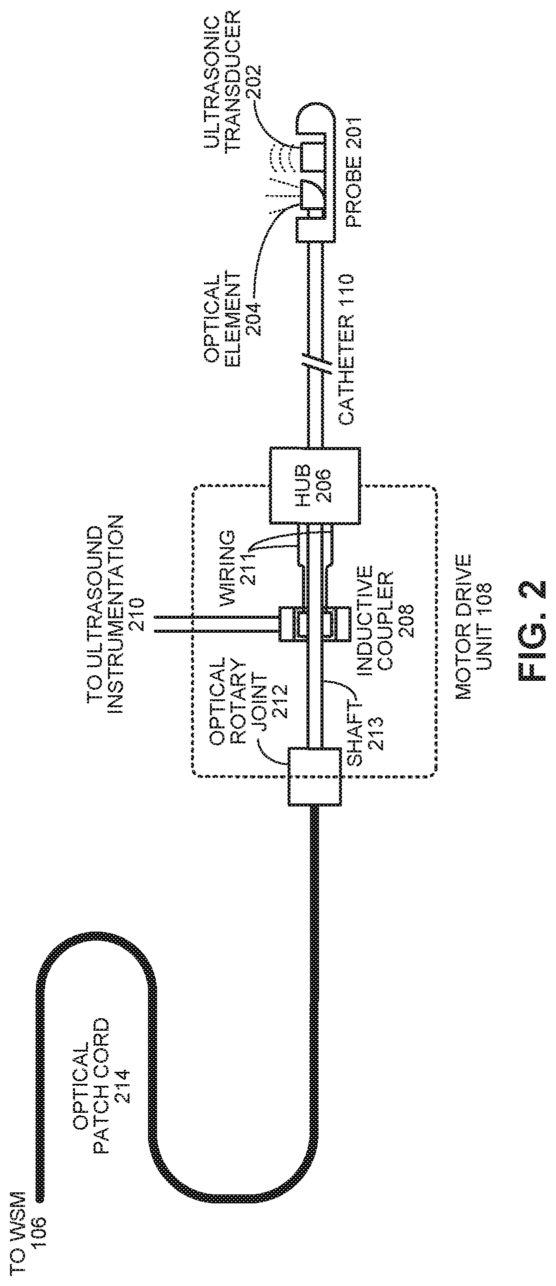
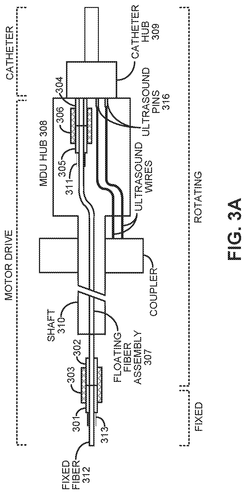
A multimodal intravascular catheter system includes a catheter with an optical channel and an electrical channel. A distal end of the catheter includes an optical element and an ultrasonic transducer, which are oriented orthogonally to a rotational axis of the catheter. A motor drive unit (MDU) is coupled to a proximal end of the catheter and includes a drive motor to rotate the catheter. The optical channel directs light from a pulsed UV laser source to the optical element, and returns an optical fluorescence signal from the optical element. A photodetector converts the returned optical fluorescence signal into an electrical fluorescence signal. An intravascular ultrasound (IVUS) processor is coupled to the ultrasonic transducer through the electrical channel, wherein the IVUS processor generates a drive signal for the ultrasound transducer, and processes echo information returned from the ultrasound transducer. Finally, a digitizer samples the electrical fluorescence signal and associated echo information.

Description

RELATED APPLICATION[0001]This application claims priority to U.S. Provisional Application No. 62 / 428,978, entitled “Apparatus and Methods for Intravascular Ultrasound Imaging and Fluorescence Lifetime Imaging,” by inventors Laura Marcu, Julien Bec and Diego Yankelevich, the contents of which are incorporated by reference herein in their entirety.GOVERNMENT LICENSE RIGHTS[0002]This invention was made with Government support under Grant No. RO1 HL067377 awarded by the National Institutes of Health. The government has certain rights to the invention.BACKGROUNDField[0003]The disclosed embodiments generally relate to catheter-based vascular-imaging techniques. More specifically, the disclosed embodiments relate to the design of a vascular-imaging system, which integrates both intravascular ultrasound (IVUS) imaging and fluorescence-lifetime imaging microscopy (FLIm) functionality into a single catheter.Related Art[0004]The majority of sudden acute cardiovascular events result from ruptur...

Claims


Application Information

Patent Timeline
Patent Type & Authority Patents(United States)
IPC IPC(8): A61B8/12A61B5/00A61B8/08A61B8/00G01N21/63A61B5/02
CPCA61B8/12A61B5/02007A61B5/6852A61B8/0891A61B5/0035A61B5/0071A61B5/6876A61B8/4444A61B8/5261G01N21/63A61B5/0062
Inventor MARCU, LAURABEC, JULIENYANKELEVICH, DIEGO R.
Owner RGT UNIV OF CALIFORNIA
Learn More