
Method of performing magnetic resonance imaging and a magnetic resonance apparatus

a magnetic resonance imaging and magnetic resonance technology, applied in the direction of nmr measurement, instruments, magnetic measurement, etc., can solve the problems of only generating t1 maps, and affecting the accuracy of mr imaging

Active Publication Date: 2021-12-07
SIEMENS HEALTHCARE GMBH +1
View PDF3 Cites 0 Cited by
  • Summary
  • Abstract
  • Description
  • Claims
  • Application Information

AI Technical Summary

Benefits of technology

The present invention aims to improve MR imaging techniques by providing a better way to create T1 maps. The technical effect of this invention is to offer an alternative approach to generate T1 maps, which may provide better accuracy and reliability in MR imaging analysis.

Problems solved by technology

The MOLLI, SASHA, and SAPPHIRE sequences are desired to be performed during a single breath-hold to reduce respiratory motion artifacts, but this can be challenging depending on the condition of the subject being imaged.
The total time required to perform the sequence, wait for the magnetization effects to return to equilibrium, and repeat the sequence is sufficiently long that it is impractical for multiple T1 maps of different regions of the subject to be generated during a single breath-hold.
As a result, the acquisition of multiple T1 maps for different regions of the subject using these existing approaches, and especially the MOLLI approach, may take a long time to be performed, and require subject cooperation to minimize the effects of motion between breath-holds.
However, these existing approaches are only capable of generating a T1 map for a single region of interest in the subject, e.g. a single slice.
The amount of time required for the magnetization to recover to equilibrium means that these existing approaches are still impractical for multiple T1 maps of different regions of the subject to be generated during a single breath-hold.
However, the robustness of these techniques is still generally inferior to breath-hold acquisitions and still require a large number of images for T1 map reconstructions (>5 images per slice) which results in long imaging protocols.

Method used


Image

  • Method of performing magnetic resonance imaging and a magnetic resonance apparatus
  • Method of performing magnetic resonance imaging and a magnetic resonance apparatus
  • Method of performing magnetic resonance imaging and a magnetic resonance apparatus

Examples


Embodiment Construction

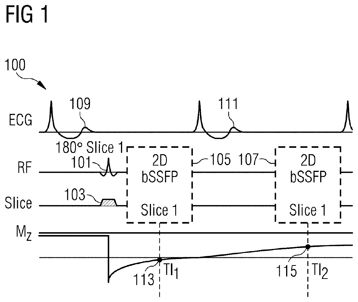
[0088]Referring to FIG. 1, there is shown a MRI pulse sequence 100 for generating a T1 map in accordance with aspects of the present invention. The sequence 100 is used for myocardial T1 mapping and is ECG gated / triggered. It will be appreciated that ECG gating is not required in all embodiments of the invention.

[0089]The sequence starts by generating a spatially selective preparation pulse 101, 103 for exciting a region of interest of the subject. The generation of the spatially selective preparation pulse 101, 103 is triggered by the ECG pulse 109. The spatially selective preparation pulse comprises an excitation pulse 101 which is an inversion pulse 101 in this example implementation and a magnetic field gradient 103 which is generated along a first spatial axis. In this example implementation, the magnetic field gradient 103 is generated along the slice select axis, and is thus a slice selective magnetic field gradient 103.

[0090]Following the generation of the spatially selectiv...


PUM


Abstract

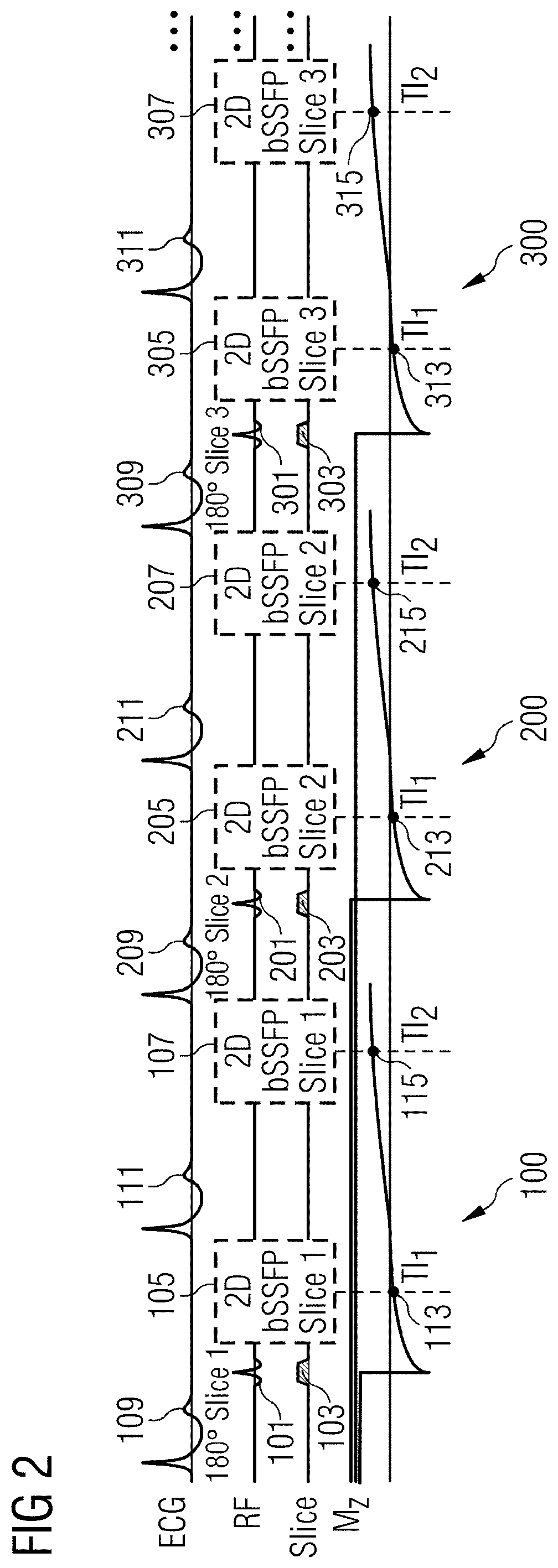
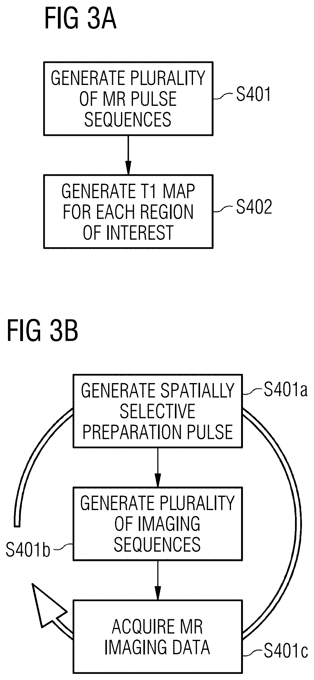
In a method and apparatus for performing magnetic resonance (MR) imaging for generating multiple T1 maps of separate regions of interest of a subject along a first spatial axis, multiple MR pulse sequences are generated, each MR pulse sequence being for imaging a respective one of the separate regions of interest of the subject. In order to generate each of the plurality of MR pulse sequences, a spatially selective preparation pulse is generated exciting the region of interest of the subject and a number of imaging sequences that follow the application of the spatially selective preparation pulse are generated. MR imaging data are acquired during the generation of the multiple imaging sequences. The multiple MR pulse sequences are generated during a period not exceeding 30 seconds.

Description

BACKGROUND OF THE INVENTIONField of the Invention[0001]The present invention relates to a method of performing magnetic resonance (MR) imaging and a magnetic resonance apparatus. In particular, the present invention relates to a method of performing magnetic resonance imaging and a magnetic resonance apparatus for generating a plurality of T1 maps.Description of the Prior Art[0002]T1 relaxation time, also known as the spin-lattice or longitudinal relaxation time, is a measure of how fast the nuclear spin magnetization returns to its equilibrium state after an excitation pulse. T1 is a key source of soft tissue contrast in MRI.[0003]Generating a T1 map of an examination area is a useful tool for a medical professional to identify diseases, lesions and abnormalities in soft tissue that affect T1. T1 mapping is useful to characterize diffuse or infiltrative myocardial diseases, including cardiomyopathies, myocarditis, and amyloidosis.[0004]T1 mapping, and in particular cardiac T1 mappi...

Claims


Application Information

Patent Timeline
Patent Type & Authority Patents(United States)
IPC IPC(8): G01R33/50G01R33/483G01R33/44G01R33/56
CPCG01R33/4838G01R33/443G01R33/4835G01R33/50G01R33/5602G01R33/56509G01R33/5673G01R33/5676G01R33/4833G01R33/4836G01R33/561G01R33/5614
Inventor HUANG, LINEJI, RADHOUENEROUJOL, SEBASTIEN
Owner SIEMENS HEALTHCARE GMBH
Learn More