Spatial index creation for IHC image analysis

a technology of image analysis and spatial index, applied in image enhancement, medical/anatomical pattern recognition, instruments, etc., can solve the problems of large computational resources, cpu and memory, large computational resources, and large number of digital images for diagnostic, educational, research purposes, and achieve the effect of quick density calculation

Active Publication Date: 2020-11-03
F HOFFMANN LA ROCHE INC
View PDF4 Cites 0 Cited by
  • Summary
  • Abstract
  • Description
  • Claims
  • Application Information

AI Technical Summary

Benefits of technology

[0013]Generating at least one spatial index for the spatial data records of one or more images of a tissue sample may be advantageous as it may now be possible to analyze spatial information of a plurality of biomedical features in a large number of digital IHC images, including whole slide images, in a highly efficient manner. Instead of using an application program for performing the analysis of the relative position of biomedical features, optimized spatial database operations are used that were originally designed for processing geodata, i.e., a completely different kind of data. Thus, data traffic to and from an application program for determining the relative position of different biomedical features in an image can be avoided. Moreover, by using spatial database operations on spatial data objects representing biomedical features instead of user-defined application program routines, speed-optimized routines that are adapted to quickly process a large number of spatial data objects can be used, thereby further reducing CPU and memory consumption.
[0072]A “spatial index” as used herein is a data structure used by spatial databases to optimize spatial queries. Spatial index types typically handle spatial queries (such as how far two points differ, or whether points fall within a spatial area of interest, whether two polygons specify disjoint, intersecting, overlapping or touching areas) more efficiently than conventional indices.

Problems solved by technology

The processing of large numbers of digital images, in particular high resolution whole slide images, for diagnostic, educational, and research purposes may be highly complex and may consume a large amount of computational resources, in particular CPU and memory.
Gains in computer processing power, data transfer speeds and cloud storage solutions have enabled the use of digital images for a wider variety of purposes in pathology, but still the processing and analysis of large numbers of digital images, in particular whole slide images, is a technological challenge that can often not be adequately solved with state of the art image processing techniques at reasonable costs.

Method used


Image

  • Spatial index creation for IHC image analysis
  • Spatial index creation for IHC image analysis
  • Spatial index creation for IHC image analysis

Examples


Embodiment Construction

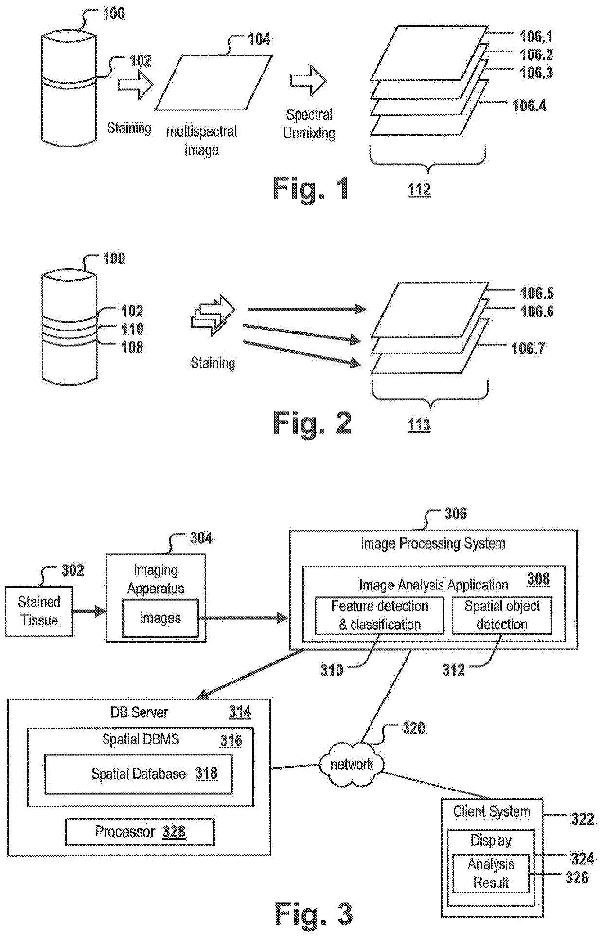
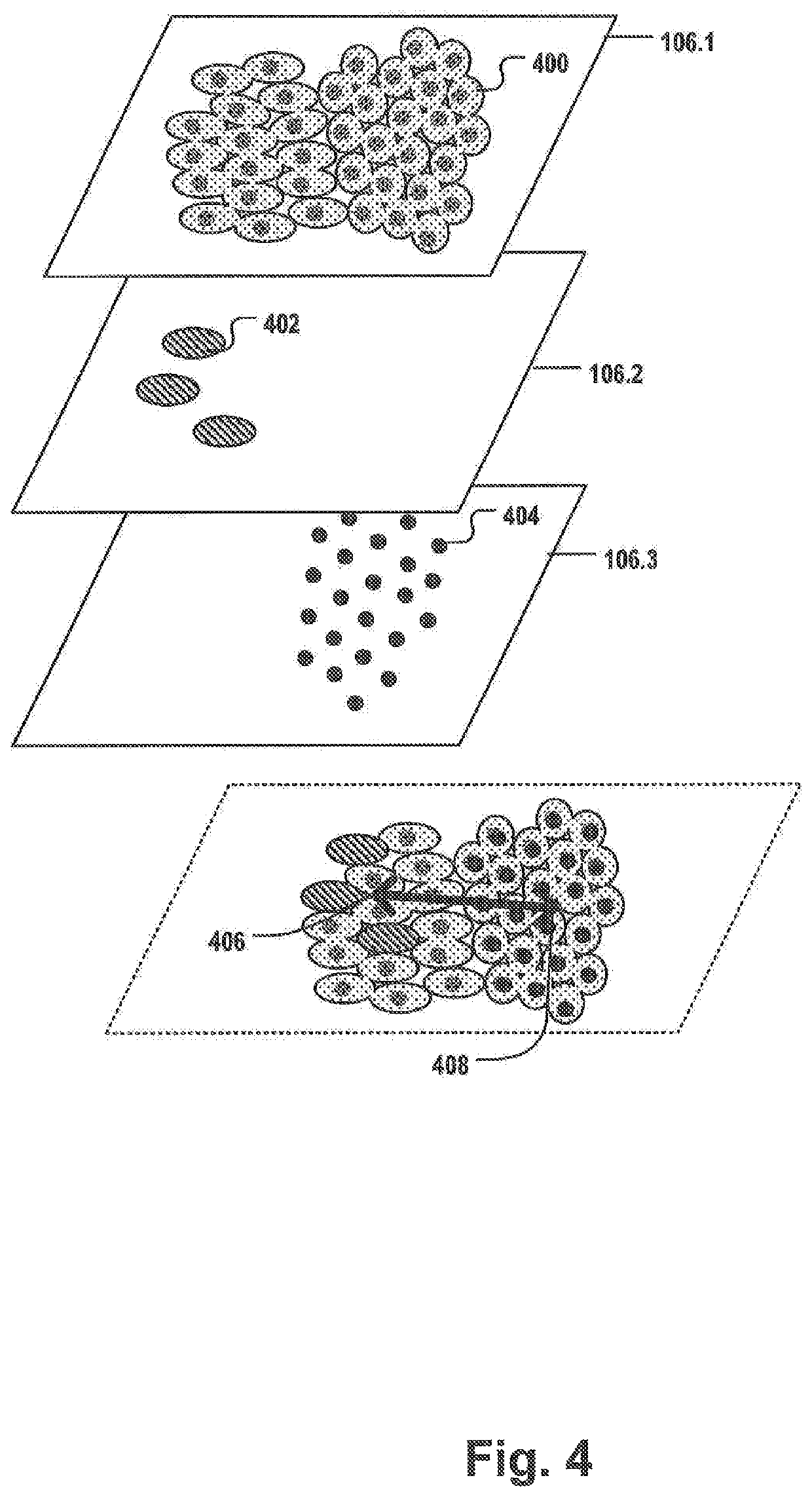
[0085]FIG. 1 shows the creation of a set 112 of digital images 106.1-106.4 of the same layer 102 of a tissue sample 100. For example, as part of the diagnosis of many cancer types, e.g. colorectal cancer, one or more biopsy samples are taken. The biopsy sample is sliced into one or more thin tissue layers 102. The layer 102 may be stained with one or more stains selectively staining specific biomarkers, cells and / or organelles, and a multispectral image 104 is taken from one layer 102 to capture meaningful biomedical features that may allow to classify the tumor, provide a prognosis and / or a treatment suggestion. The multispectral image may comprise spectral information of a plurality of different stains, e.g. fluorescent stains, and / or may cover a whole slide comprising the layer 102. Thus, the resulting multispectral image is often very large. By applying a spectral unmixing procedure, a plurality of digital images 106-1-106.4 is created. Each of the images in set 112 may correspo...


PUM


Abstract

An image analysis method for analyzing an IHC tissue sample includes storing, for each of one or more sets of digital images, a set-ID in a spatial database of a spatial DBMS, the images of each set depicting the same or adjacent layers of the IHC tissue sample; storing, for each of the, a plurality of spatial data objects in the database in association with the set-ID; creating, by the spatial DBMS, at least one spatial index covering the objects of the images contained in the one or more sets; applying, by the spatial DBMS, a spatial database operation on the at least one spatial index for determining the relative positions of objects of two or more images of a selected set; and providing the relative positions as input for determining the distance and / or relative position of the biomedical features of the tissue sample.

Description

CROSS-REFERENCE TO RELATED APPLICATIONS[0001]This application is a national phase under 35 U.S.C. § 371 of PCT International Application No. PCT / EP2017 / 054917 which has an International filing date of Mar. 2, 2017, which claims priority to European Application No. 16164102.2, filed Apr. 6, 2016, the entire contents of each of which are hereby incorporated by reference.FIELD OF THE INVENTION[0002]The present invention relates to an image analysis system for analyzing images of IHC tissue samples.BACKGROUND AND RELATED ART[0003]A plurality of digital imaging solutions in pathology exist aiming at generating a large number of digital images of tissue samples and / or generating high-resolution images of whole slides. The processing of large numbers of digital images, in particular high resolution whole slide images, for diagnostic, educational, and research purposes may be highly complex and may consume a large amount of computational resources, in particular CPU and memory. Gains in com...

Claims


Application Information

Patent Timeline
Patent Type & Authority Patents(United States)
IPC IPC(8): G06T7/73G06K9/00G06K9/62G06T7/60G06F16/51G06T7/00G16H30/20G06T7/30G06T7/50
CPCG06T7/0012G06T7/73G06K9/00147G16H30/20G06K9/6212G06T7/30G06F16/51G06T7/50G06K9/0014G06T7/60G06T2207/30024G06T2207/30028G06K2209/05G06T2207/30096G06V20/695G06V20/698G06V2201/03
Inventor MECH, FRANZISKAFUCHS, ANGELIKAKUENTZER, JANHUBER, OTTOKOHN, ALEXGUTHOEHRLEIN, ECKHART
Owner F HOFFMANN LA ROCHE INC
Why Eureka
Try Eureka