
Confocal laser fundus angiographic device

Active Publication Date: 2020-03-03
SUZHOU MICROCLEAR MEDICAL INSTR
View PDF12 Cites 1 Cited by
  • Summary
  • Abstract
  • Description
  • Claims
  • Application Information

AI Technical Summary

Benefits of technology

The device ensures accurate and reliable filter switching, preventing imaging failures by automating the process and ensuring the correct filter is engaged for each imaging mode, thereby improving diagnostic accuracy and reducing operator error.

Problems solved by technology

The operator is likely to forget to switch the filter or switch the filter incorrectly due to a mistake, resulting in imaging failure.

Method used


Image

  • Confocal laser fundus angiographic device
  • Confocal laser fundus angiographic device
  • Confocal laser fundus angiographic device

Examples


Embodiment Construction

[0023]In order to clear the purpose, the technical scheme and the advantages of the invention, reference will now be made to the drawing figures to describe the embodiments of the present disclosure in detail.

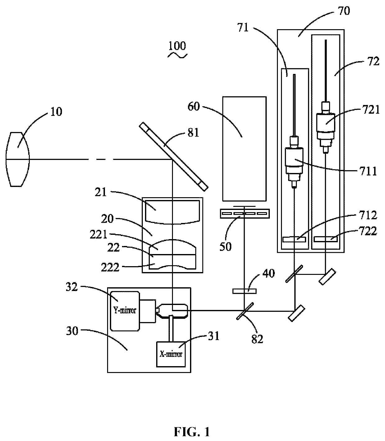
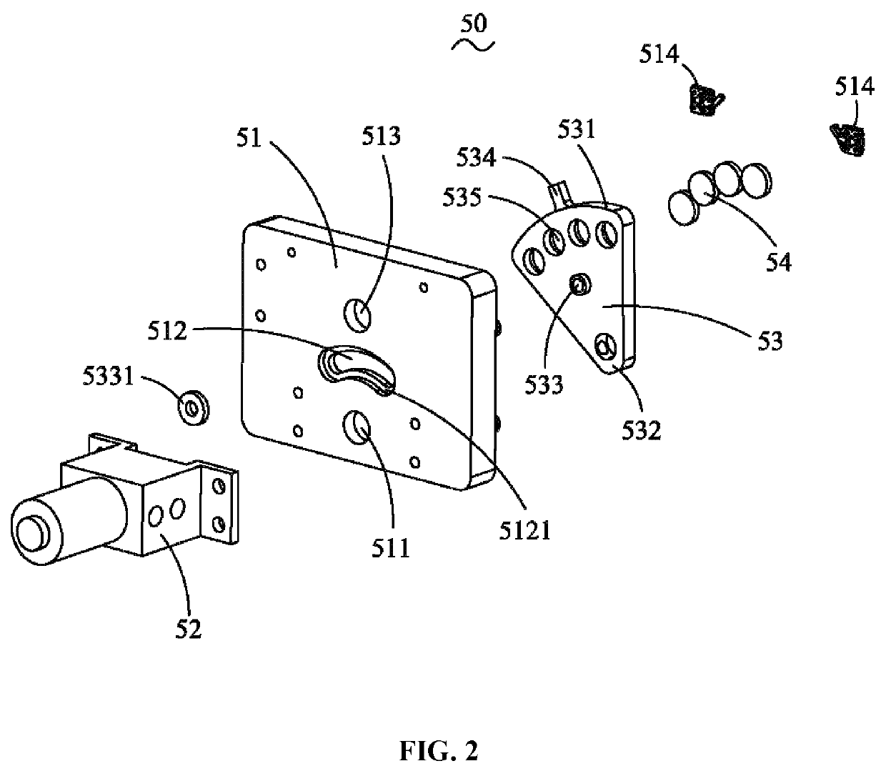
[0024]Referring to FIG. 1, the present invention discloses a confocal laser fundus angiographic device 100 comprises an objective lens 10, a scanning lens 20, a scanning galvanometer 30, a mirror 40, a filtering module 50, an imaging detection assembly 60 and excitation light source 70. The excitation light source 70 emits illumination light passes through the scanning galvanometer 30, the scanning lens 20, the objective lens 10, and then enters in the fundus, the fluorescent substances in the fundus vein are excited by the illumination light to emit light with a specific wavelength, the specific wavelength light passes through the objective lens 10, the scanning lens 20, the scanning galvanometer30, the mirror 40, the filtering module 50, and then enters in the imaging detecti...


PUM

PropertyMeasurementUnit
wavelengthaaaaaaaaaa
specific wavelengthaaaaaaaaaa

Abstract

A confocal laser fundus angiographic device, comprises: an objective lens, a scanning lens, a scanning galvanometer, a mirror, a filtering module, an imaging detection assembly and an excitation light source. The illumination light emitted by the excitation light source enters the fundus passing through the scanning galvanometer, the scanning lens and the objective lens. Fluorescent substances in fundus vessels are excited by the illumination light and emit light with a specific wavelength. The light with a specific wavelength enters the imaging detection assembly by passing through the objective lens, the scanning lens, the scanning galvanometer, the mirror and the filtering module. The filtering module comprises a base, a motor mounted on the base, a rotary block mounted on the motor and at least one filter mounted on the rotary block. The base is provided with a light-through hole. Compared with the prior art, the confocal laser fundus angiographic device is capable of automatically switching the filter, thereby avoiding the problem of forgetting to switch the filter due to the operator's mistake or switching the filter by mistake.

Description

CROSS-REFERENCE TO RELATED APPLICATIONS[0001]This application claims priority to Chinese Application Serial No. 201610775302.5, filed Aug. 31, 2016, the disclosures of which are hereby incorporated by reference in their entirety.TECHNICAL FIELD[0002]The present invention generally relates to angiography imaging, and more particularly to a confocal laser fundus angiographic device.BACKGROUND[0003]Many eye diseases are associated with fundus changes, such as age-related macular degeneration (ARMD), diabetic retinitis, intraocular tumors, central serous chorioretinopathy (CSC), harada's disease and so on. Fundus fluorescein angiography and indocyanine green angiography are important diagnostic technique to inspect and record the condition of retina and choroidal vascular. In the 1960s, modern fundus fluorescein angiography began to be used in clinical medicine, and the indocyanine green angiography began to apply in clinical medicine in the mid-90s. It is of great significance to obtai...

Claims


Application Information

Patent Timeline
Patent Type & Authority Patents(United States)
IPC IPC(8): A61B3/12A61B3/00A61B3/15A61B3/10
CPCA61B3/156A61B3/0008A61B3/1025A61B3/12A61B3/1241A61B3/14A61B3/15
Inventor LI, LEIZHAO, ENYIHOU, XIAOPINGLI, CHAOHONG
Owner SUZHOU MICROCLEAR MEDICAL INSTR
Learn More