Methods and compositions for preparing samples for immunostaining
a technology of immunostaining and composition, which is applied in the field of immunostaining preparation methods and compositions, can solve the problems of degrading the cellular morphology of cytology specimens, high heat pretreatment, etc., and achieves the effect of maintaining cellular morphology
- Summary
- Abstract
- Description
- Claims
- Application Information
AI Technical Summary
Benefits of technology
Problems solved by technology
Method used
Image
Examples
example 1
of Antigen Retrieval Solutions and Incubation Temperatures
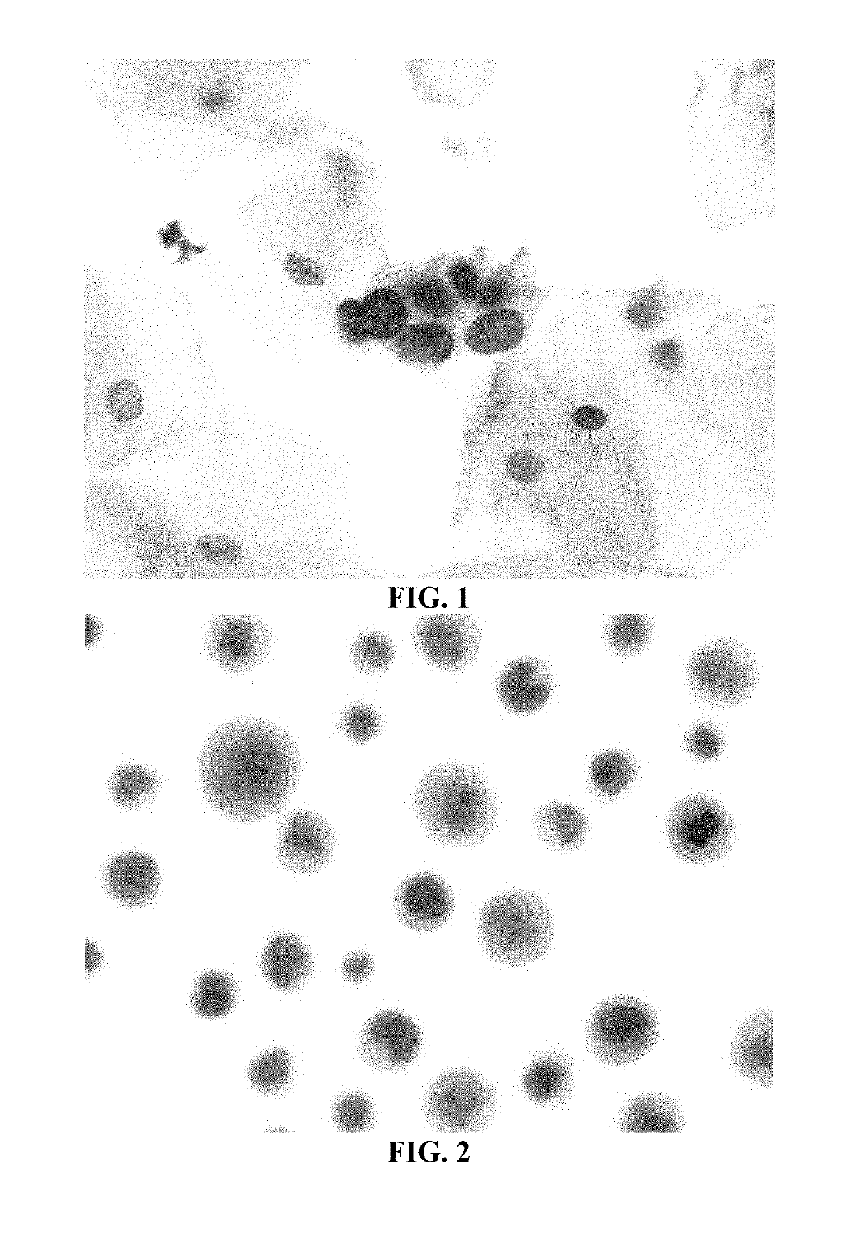
[0178]Historically, most tissue-based antigen retrieval (AR) techniques have utilized high heat, ˜100° C. and / or treatment with various solutions. The AR procedure for the detection of the antigens MCM2 and MCM7 in SurePath® (TriPath Imaging, Inc.) cervical cytology samples using a cocktail of three antibodies disclosed in U.S. Pat. Nos. 7,595,380 (27C5.6 and 26H6.19 anti-MCM2 antibodies) and 7,632,498 (2E6.2 anti-MCM7 antibody), which are herein incorporated by reference in their entirety, was optimized as described hereinbelow to maintain cell morphology, while ensuring appropriate immunostaining of cytology specimens by investigating multiple AR solutions and incubation temperatures.
[0179]A broad range of potential AR (pretreatment) solutions (n=43) were tested on SurePath® cytology specimens to determine which ones would have a positive effect on immunostaining with a triple antibody cocktail (two anti-MCM2 antibodies and...
example 2
n of the Effects of the Two-Step Pretreatment Method with a High-Heat Pretreatment Method on Immunostaining and Morphology in Cervical Cytology Samples
[0196]This study compared the two-step pretreatment method using two different chaotropic salt solutions for the second incubation step to each other and to a pretreatment method utilizing high heat (i.e., the steamer method described in Example 1).
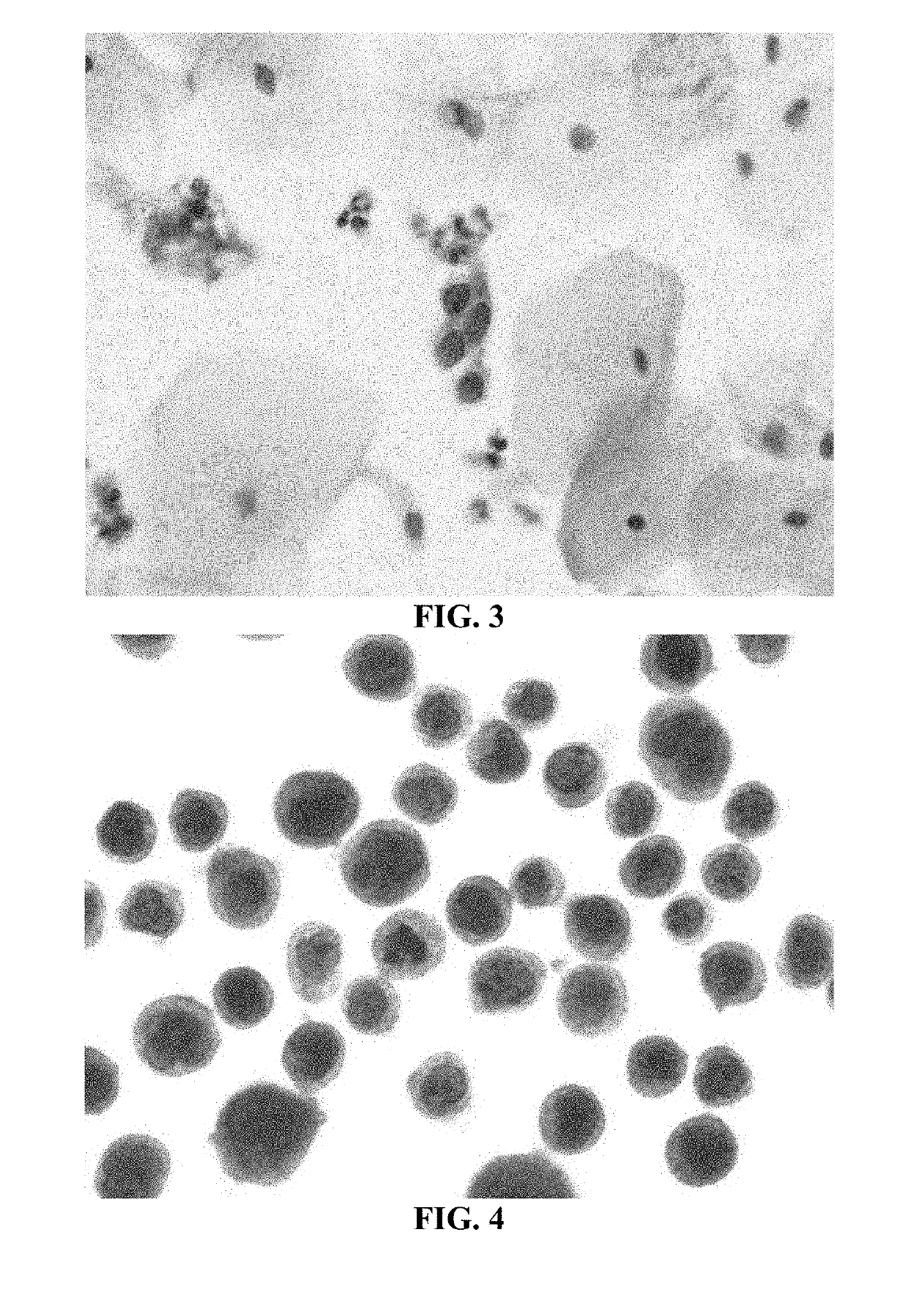
[0197]In these experiments, SurePath® cervical cytology specimens were processed using the internally developed PrepStain Plus® Instrument (Tripath Imaging, Inc.) and deposited for staining. For those samples treated with the two-step pretreatment method, the slides were heated to 50° C. on specially-designed slide heating trays and Pretreatment Solution 1 (0.1% SDS) was applied and the slides were incubated at 50° C. for 19 minutes. The cells were washed with a standard Tris-buffered saline (TBS) solution. Pretreatment Solution 2 (either 3M LiClO4 / 0.1% NP-40 or 3M guanidine thiocyanate / 0.1...
example 3
of Antigens Ki67 and p16 in Cervical Cytology Samples Using the Two-Step Pretreatment Process
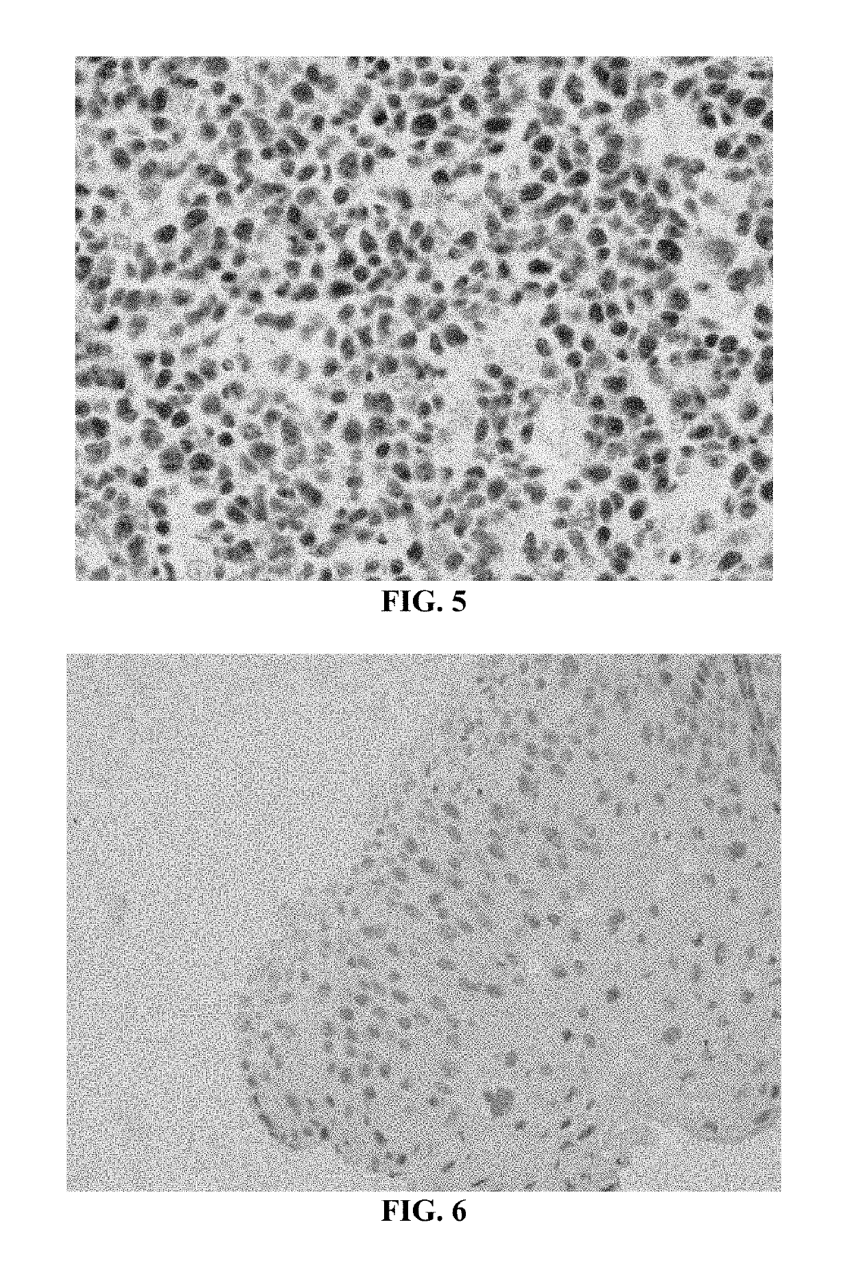
[0218]In these experiments, SurePath® cervical cytology specimens were processed using the internally developed PrepStain Plus® Instrument (Tripath Imaging, Inc.) and deposited for staining. Following cell deposition, the slides were heated to 50° C. on specially-designed slide heating trays and AR Solution 1 (0.1% SDS) was applied and the slides were incubated at 50° C. for 19 minutes. The cells were washed with a standard tris-buffered saline (TBS) solution. AR Solution 2 (3M LiClO4 / 0.1% NP-40) was then added and the cells were incubated an additional 19 minutes at 50° C. Following this incubation, the slides were washed again with TBS and processed for immunostaining and PAP counterstaining on the PrepStain Plus® instrument. The slides were then coverslipped and examined by a cytologist and / or cytopathologist. Representative results of cervical cytology samples immunostained with an anti-...
PUM
| Property | Measurement | Unit |
|---|---|---|
| temperatures | aaaaa | aaaaa |
| pH | aaaaa | aaaaa |
| temperature | aaaaa | aaaaa |
Abstract
Description
Claims
Application Information
 - R&D
- Intellectual Property
- Life Sciences
- Materials
- Tech Scout
- Unparalleled Data Quality
- Higher Quality Content
- 60% Fewer Hallucinations
Browse by: Latest US Patents, China's latest patents, Technical Efficacy Thesaurus, Application Domain, Technology Topic, Popular Technical Reports.
© 2025 PatSnap. All rights reserved.Legal|Privacy policy|Modern Slavery Act Transparency Statement|Sitemap|About US| Contact US: help@patsnap.com


