
In vitro detection of prions in blood

a technology of prions and in vitro detection, applied in the field of infectious agent screening and diagnosis, can solve the problems of many months, animals, cost, etc., and achieve the effects of low prpd, rapid identification and diagnosis, and sufficient sensitivity

Active Publication Date: 2019-07-23
COLORADO STATE UNIVERSITY
View PDF2 Cites 1 Cited by
  • Summary
  • Abstract
  • Description
  • Claims
  • Application Information

AI Technical Summary

Benefits of technology

[0009]The present invention provides extremely sensitive methods for detecting prion protein, including PrPres or PrPSc, in a sample The modified rPrPD amplification assays are surprisingly much faster than existing PMCA methods, yet it still retains sufficient sensitivity to detect extremely low levels of PrPD. The method permits the rapid identification and diagnosis of prion disease, which can limit the transmission of prion diseases, particularly through the blood supply.
[0010]Blood-borne transmission of infectious prions during the asymptomatic or pre-clinical stage of disease occurs for both human and animal transmissible spongiform encephalopathies (TSEs). The geographical distribution of the cervid TSE, chronic wasting disease (CWD), continues to spread across North America and the prospective number of individuals harboring an asymptomatic infection of human variant Creutzfeldt-Jakob Disease (vCJD) in the United Kingdom has been projected to be ˜1 in 3000 residents. Thus, it is important to monitor cervid and human blood products to ensure herd health and human safety. Current methods for detecting blood-associated prions rely primarily upon bioassay in laboratory animals. While bioassay provides high sensitivity and specificity, it requires many months, animals, and costs. Modifications of the real time quaking-induced conversion (RT-QuIC) assay enables detection of blood-borne prions in whole blood from prion-infected preclinical white-tailed deer, muntjac deer, and Syrian hamsters, attaining sensitivity of >90% while maintaining 100% specificity. Our results indicate that RT-QuIC methodology as modified and taught herein can provide consistent and reliable detection of blood-borne prions in pre-clinical and symptomatic stages of two animal TSEs, offering promise for prionemia detection in other species, including humans.
[0019]In an advantageous embodiment the method can include the step of screening the amplified PrPD for the presence of PrPD. In other words, it will often be unknown whether a particular sample contains PrPD. By virtue of the amplification, the PrPD in the sample, if any, should have been increased to detectable levels, where it would not have been detectable prior to amplification. The absence of PrPD in the sample upon attempted detection after amplification would indicate that the original sample was free of PrPD. In a similar manner, the method can include the step of contacting the sample with thioflavin T (ThT) prior to an incubating step and measuring the fluorescence of the resulting sample after an incubating step. The fluorescence of the ThT in the sample following incubation allows the presence of PrPD in the sample to be determined. More particularly, the fluorescence emitted by the sample increases upon the transition of PrPC to PrPD. Therefore, an increase in the fluorescence will correlate with an increase in the amount of PrPin the sample.
[0020]In a second aspect the present invention provides a method for the detection of a prion disease-associated conformer of prion protein (PrPD) in a sample. The method of the second aspect includes the steps of providing a sample to be screened for PrPD; freezing and thawing the sample; contacting the thawed sample with an excess of a non-pathogenic conformer of PrPC or a non-pathogenic conformer of recombinant PrP; incubating the contacted sample with the non-pathogenic conformer; disaggregating any aggregates of PrPD formed during the incubating step; repeating the incubating and disaggregating steps one or more times to yield an amplified PrPD in the sample; and screening the incubated sample for PrPD. The method of the second aspect amplifies the PrPD in the sample, if any, to detectable levels. In an advantageous embodiment the method can include the steps of contacting the sample with ThT prior to an incubating step and measuring the fluorescence of the resulting sample after an incubating step. The level of fluorescence of the ThT in the sample following incubation allows the presence of PrPD in the sample to be determined.

Problems solved by technology

While bioassay provides high sensitivity and specificity, it requires many months, animals, and costs.

Method used


Image

  • In vitro detection of prions in blood
  • In vitro detection of prions in blood
  • In vitro detection of prions in blood

Examples


example 1

nalysis of Whole Blood Collected in Various Anticoagulants

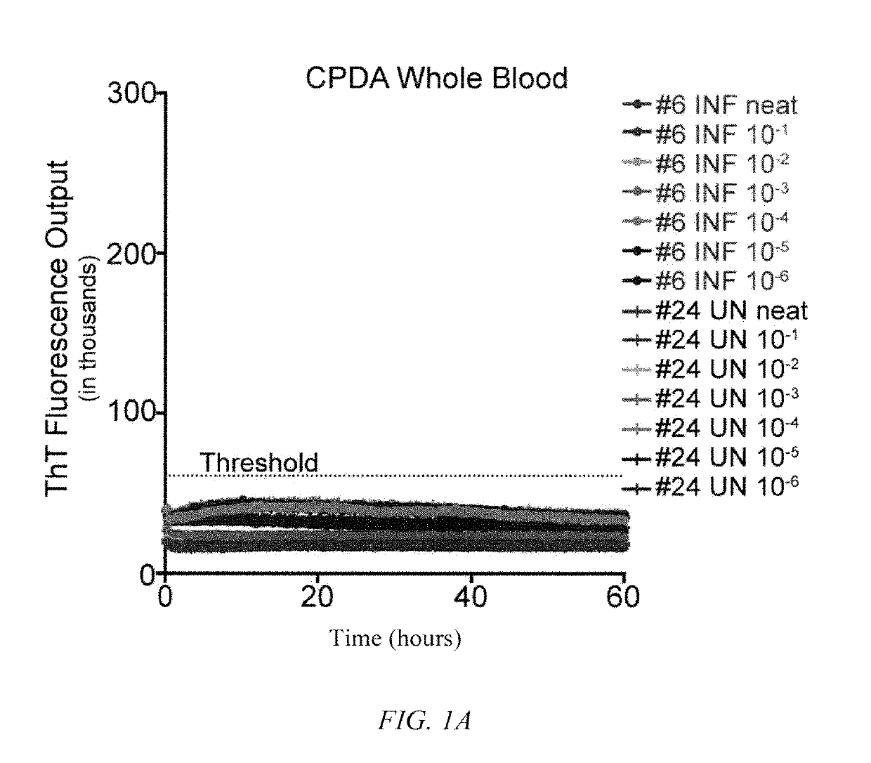
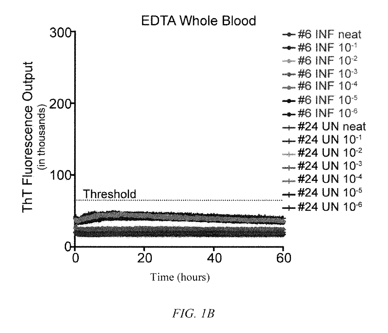
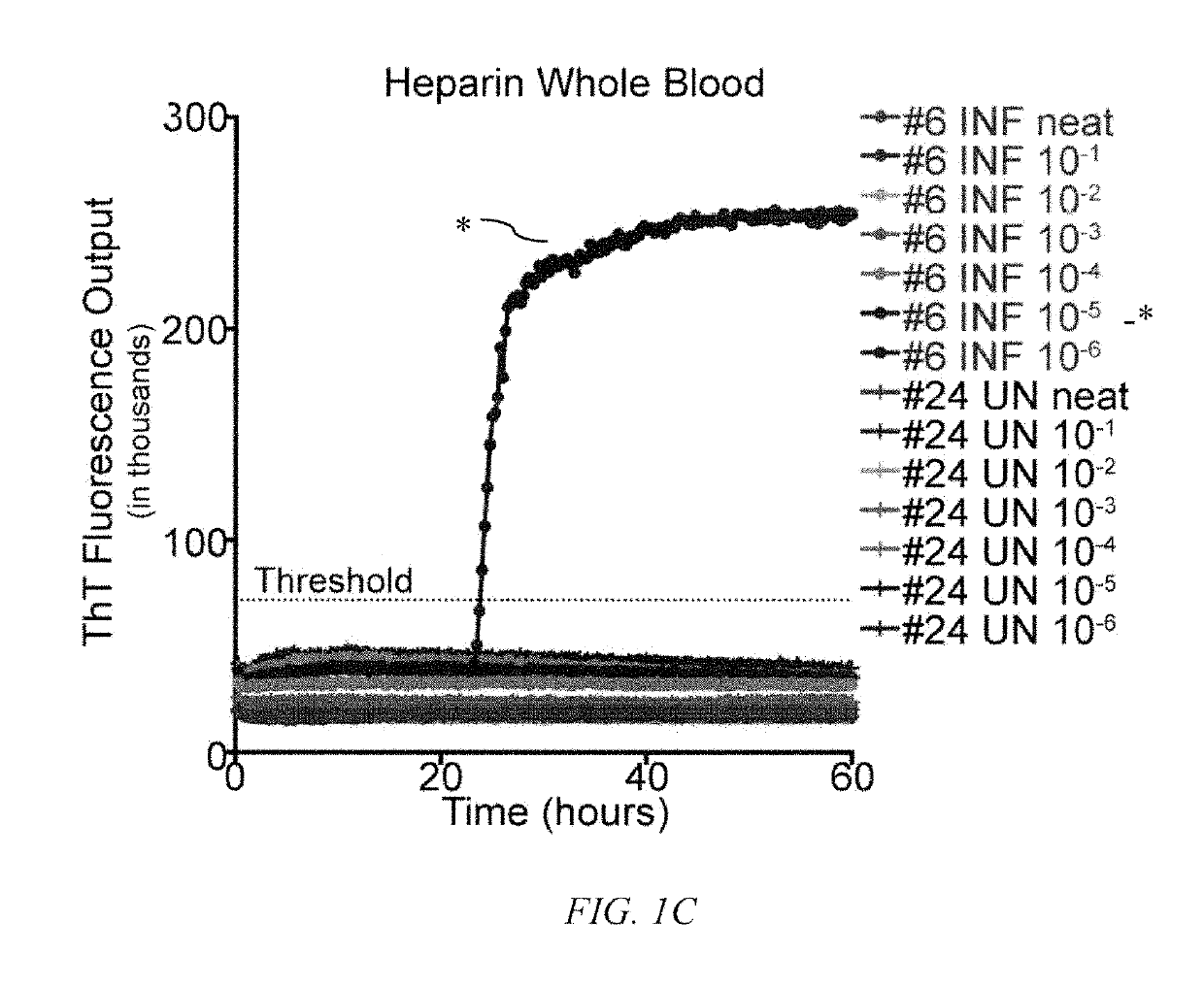
[0062]To determine the influence of common blood preservation reagents in in vitro PrPD detection assays, we compared the ability of RT-QuIC to amplify CWD prions in cervid whole blood preserved in CPDA, EDTA or heparin. Samples were run in serial dilutions (100-10−6) in the RT QuIC assay to determine the optimal dilution for PrPD detection. While RT-QuIC PrPC converting activity was observed in heparinized blood from CWD-infected deer (½ replicates in one dilution; 10−5) (FIG. 1C), PrPC converting activity was not detected in CPDA (FIG. 1A) or EDTA (FIG. 1B) preserved blood from the same animal or any blood collected from sham-inoculated deer (FIGS. 1A-1C). All subsequent RT-QuIC analysis was conducted on whole blood harvested in heparin.

[0063]Precedence for hematogenous spread of prions via transfusion has been well established with various TSEs, including scrapie [Andreoletti, O., et al., PLoS Pathog, 2012. 8(6): p. e10027...

example 2

nalysis of Fresh Versus Frozen Whole Blood

[0065]In order to determine if historical blood samples were adequately preserved to initiate PrPC converting activity in RT-QuIC, whole blood was collected from contemporary naïve and CWD112 infected white-tailed deer and compared as fresh versus frozen samples. Samples were processed in various dilutions ranging from undiluted to 10−6 to determine the optimal dilution for PrPD detection using frozen whole blood in the RT-QuIC assay. While PrPC converting activity was detected in fresh whole blood (FIG. 2A), blood that had been processed through the freeze-thaw procedure yielded higher and more consistent detection of prion converting activity (2 / 2 replicates in each of four dilutions) (FIG. 2B). PrPC converting activity was not observed in wells containing only substrate or naïve cervid blood. All subsequent RT-QuIC analysis included heparinized whole blood that had undergone four freeze-thaw cycles.

[0066]To assess the feasibility of using...

example 4

omparison of CWD-Positive Brain Versus NaPTA Concentrated Whole Blood

[0071]To evaluate the levels of PrPD present in NaPTA concentrated whole blood samples, PrPC converting activity was compared to that detected in serial dilutions of CWD-positive white tailed deer brain (FIG. 4). NaPTA treated whole blood (10 ml starting volume of whole blood) diluted to 10−2 demonstrated PrPD levels approximately equivalent to that measured in 10−6-10−7 dilution of CWD-positive brain. Equivalence was determined by comparison of the time to positivity for whole blood and brain samples.

[0072]Many groups have developed quantitative in vitro methodologies to analyze the levels of PrPD present in various tissues and bodily fluid samples. Murayama et al. [Murayama, Y., et al., J Gen Virol, 2007. 88(Pt 10): p. 2890-8.] used PMCA to establish a direct comparison of PrPD levels in buffy coat and plasma to PrPD levels seen in serial dilutions of TSE-infected brain by analyzing which round of PMCA samples be...


PUM

PropertyMeasurementUnit
temperatureaaaaaaaaaa

Abstract

A method of screening a blood sample for the presence of prions. The method includes the steps of collecting the blood sample in heparin, contacting the sample with a solution comprising recombinant prion protein (rPrP) and Thioflavin T (ThT), and measuring the resulting ThT fluorescence in the sample. The method can further include the step of freezing and thawing the sample prior to contacting the sample with a solution comprising recombinant prion protein (rPrP) and Thioflavin T (ThT). The method can also include the step of precipitating the prions in sodium phosphotungstic acid (NaPTA) prior to contacting the sample with a solution comprising recombinant prion protein (rPrP) and Thioflavin T (ThT).

Description

CROSS-REFERENCE TO RELATED APPLICATIONS[0001]This application is a continuation-in-part of U.S. application Ser. No. 14 / 920,422, filed Oct. 22, 2015, which claims the benefit of U.S. Provisional Application No. 62 / 067,016, filed Oct. 22, 2014.STATEMENT OF GOVERNMENT INTEREST[0002]This invention was made with government support under Grant Nos. R01 NS061902, N01 AI025491 and R01 AI093634 awarded by the National Institutes of Health. The government has certain rights in the invention.FIELD OF INVENTION[0003]This invention relates to infectious agent screening and diagnoses. More specifically, this invention relates to in vitro detection of prions and infectious proteins in blood.BACKGROUND OF THE INVENTION[0004]The hematogenous spread of prions in transmissible spongiform encephalopathy (TSE)-infected animals has long been hypothesized, but evidence for the presence of prions in non-nervous / lymphoid tissues and blood was not available for several decades. Later studies have provided u...

Claims


Application Information

Patent Timeline
Patent Type & Authority Patents(United States)
IPC IPC(8): G01N33/68
CPCG01N33/6893G01N33/6896G01N2800/2828
Inventor HOOVER, EDWARD A.MATHIASON, CANDACE K.
Owner COLORADO STATE UNIVERSITY
Learn More