
Medical user interfaces and related methods of use

a user interface and medical technology, applied in the field of medical user interfaces, can solve the problems of inaccurate and/or cumbersome current methods of sizing objects in the operating field, and achieve the effects of reducing the number of users

Active Publication Date: 2019-04-16
BOSTON SCI MEDICAL DEVICE LTD
View PDF9 Cites 176 Cited by
  • Summary
  • Abstract
  • Description
  • Claims
  • Application Information

AI Technical Summary

Benefits of technology

The present patent is about a medical system for use in a lithotripsy procedure, which involves using an imaging device to capture images of a patient's kidneys, bladders, or ureters. The system includes a processor that receives the image data and analyzes it to detect the presence of an object within the image. The processor can estimate the size of the object and display it on a screen. The system can also include a second imaging device that sends x-ray data to the processor to help with the analysis. The system can also provide recommendations for inflating a balloon or delivering a stent during the procedure. The method for using the system involves analyzing the image and updating the size of the object in real-time as fragments of the object are removed. The system and method can help improve the accuracy and safety of the lithotripsy procedure.

Problems solved by technology

These stones may block ducts and cause great pain and therefore must be removed.
However, current methods of sizing objects in the operating field are inaccurate and / or cumbersome.

Method used


Image

  • Medical user interfaces and related methods of use
  • Medical user interfaces and related methods of use
  • Medical user interfaces and related methods of use

Examples


Embodiment Construction

[0015]Reference will now be made in detail to examples of the present disclosure, which are illustrated in the accompanying drawings. Wherever possible, the same reference numbers will be used throughout the drawings to refer to the same or like parts.

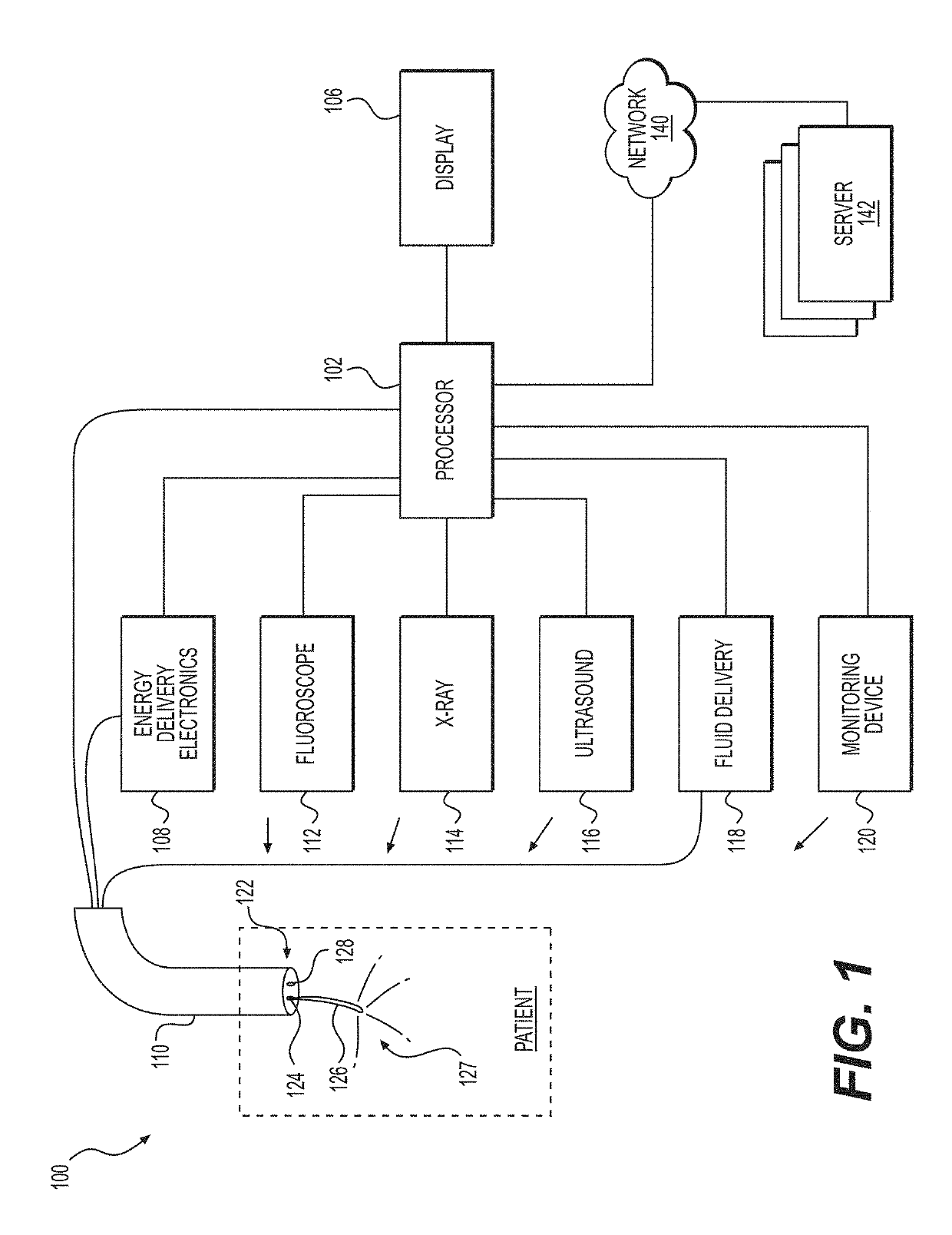
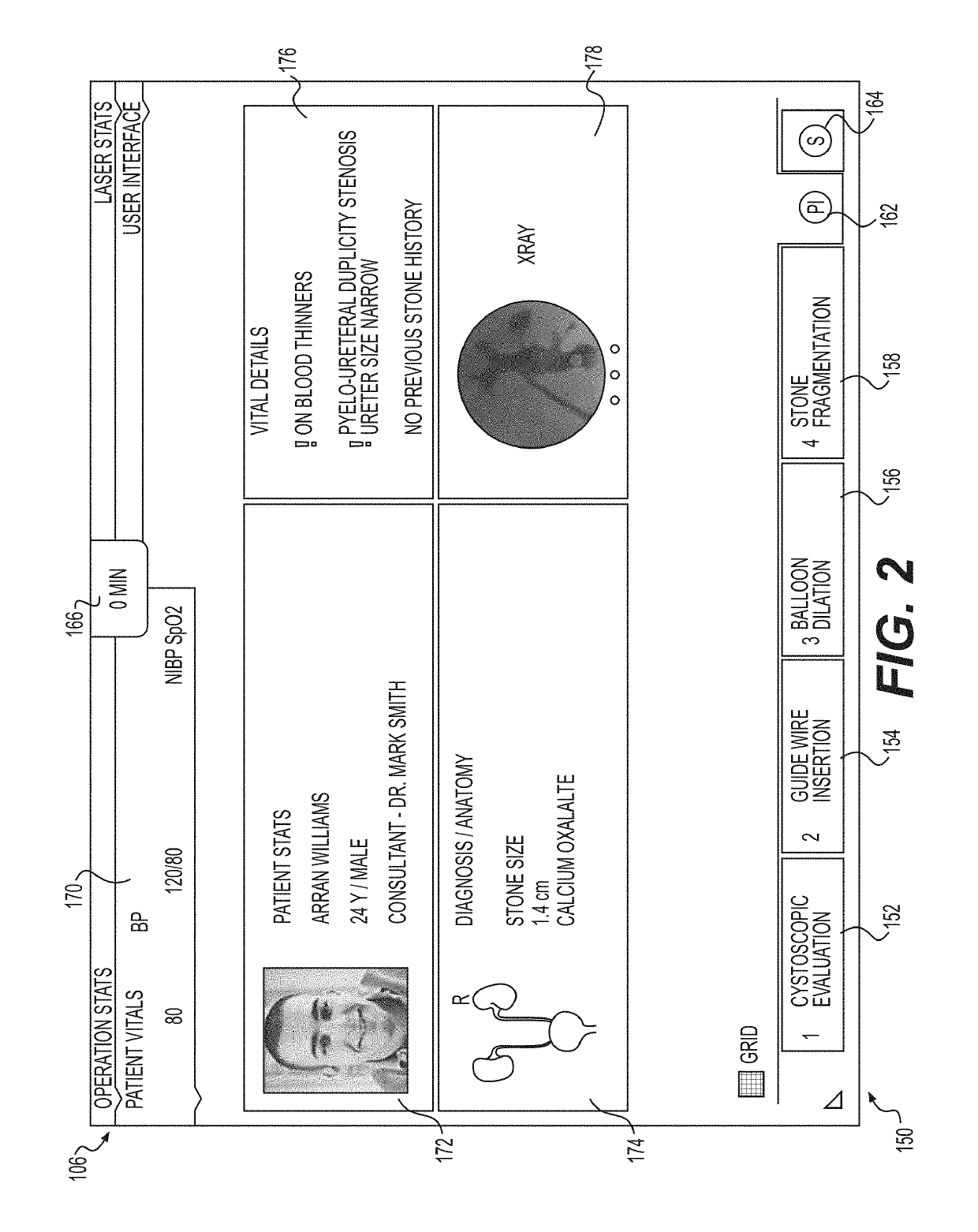
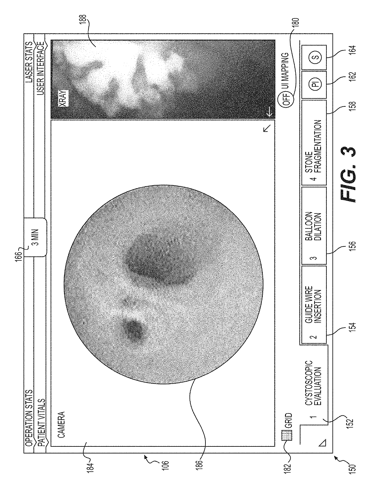
[0016]Examples of the present disclosure relate to devices and methods for controlling the application of energy to objects disposed within a body lumen of a patient, such as, e.g., a lumen of a kidney, a bladder, or a ureter. FIG. 1 illustrates a system 100 for delivering energy, in accordance with a first example of the present disclosure. The system may include a processor 102 that is operatively coupled to a display 106. In some examples, processor 102 and display 106 may be disposed within a single handheld unit, such as, e.g., a tablet computer such as a Microsoft Surface, iPAD® or iPHONE®. In other examples, processor 102 and display 106 may be modular and may connect to one another by any suitable mechanism. Display 106 may be ...


PUM


Abstract

A medical system for use in a lithotripsy procedure may include a processor configured to receive input from a first imaging device, wherein the first imaging device may be configured to send image data representative of an image captured in a lumen of a kidney, bladder, or ureter to the processor. The processor may be configured to display the image on a display device coupled to the processor, and analyze the image to sense the presence of an object within the image. If an object was sensed within the image, the processor may analyze the image to estimate a size of the object, and display the estimate on the display device.

Description

CROSS-REFERENCE TO RELATED APPLICATION(S)[0001]This patent application claims the benefit under 35 U.S.C. § 119 to U.S. Provisional Patent Application No. 62 / 288,654, filed on Jan. 29, 2016, the entirety of which is incorporated herein by reference.TECHNICAL FIELD[0002]Examples of the present disclosure relate generally to medical user interfaces and related methods of use. More particularly, examples of the present disclosure relate to medical user interfaces for use in lithotripsy procedures.BACKGROUND[0003]Many patients develop stones within their common bile, urinary, renal or ureteral systems. These stones may block ducts and cause great pain and therefore must be removed. Several approaches are available for treating such stones, including, laser treatment and subsequent removal of the stones from the body. These lithotripsy procedures are often performed with the aid of an endoscopic device, and an endoscopic operating field may be displayed to an operator. Sizing objects in ...

Claims


Application Information

Patent Timeline
Patent Type & Authority Patents(United States)
IPC IPC(8): A61B34/00A61B18/26G06T11/60G06T7/00G06T7/60G16H40/63A61B34/10A61B34/20A61B6/00A61B17/22A61B18/00
CPCA61B34/25G06T7/0014G06T7/60G06T11/60G16H40/63A61B18/26G06T2210/41A61B6/464A61B6/465A61B17/22004A61B2018/00505A61B2018/00511A61B2018/00517A61B2018/00577A61B2018/00982A61B2034/107A61B2034/2065A61B2034/2074A61B2034/252A61B2034/254A61B2034/256A61B2217/007A61B2218/002G06T2207/10116G06T2207/30084
Inventor HARRAH, TIMOTHY P.OSKIN, CHRISTOPHER L.LENZ, DERRICKBANERJEE, ARPITAGAVADE, SANDESHMISRA, PAVANTAKALE, ABHIJIT
Owner BOSTON SCI MEDICAL DEVICE LTD
Learn More