Eureka AIR delivers breakthrough ideas for toughest innovation challenges, trusted by R&D personnel around the world.

Lung volume reduction method

a lung volume and volume reduction technology, applied in medical science, surgical instruments for cooling, surgery, etc., can solve the problems of lack of versatility of the method, inability to apply the method to such a patient in whom the tissues to be raked up have been lost, and the candidate patients for application of the therapeutic method are limited, so as to achieve speedy and easy treatment. the effect of access

Active Publication Date: 2018-10-02
TERUMO KK
View PDF9 Cites 0 Cited by
  • Summary
  • Abstract
  • Description
  • Claims
  • Application Information

AI Technical Summary

Benefits of technology

[0015]According to the aspect of the present disclosure as described, it is possible to reduce the volume of the lung having an emphysema-suffering part, and to alleviate the feeling of difficulty in breathing. Therefore, it is possible to exhibit a reliable therapeutic effect on chronic obstructive pulmonary disease.
[0027]According to the aspect of the disclosure as described, the restraining of the expansion and contraction of the lung parenchyma is conducted by use of a medical device introduced transbronchially and / or a medical device introduced from a body surface side. Therefore, an introducing method promising easier access can be arbitrarily selected according to the part to be treated and the kind of the medical device, and the treatment can be carried out speedily and easily.

Problems solved by technology

This method, however, cannot be applied to such a patient in whom the tissues to be raked up have been lost in the lung parenchyma and the surroundings due to the progress of the emphysema.
Therefore, the candidate patients for application of this therapeutic method would be limited depending on the symptom of the affected part (lesion part).
Thus, this method lacks versatility.

Method used


Image

  • Lung volume reduction method
  • Lung volume reduction method
  • Lung volume reduction method

Examples


example

[0076]A working example of the present disclosure will be described herein below.

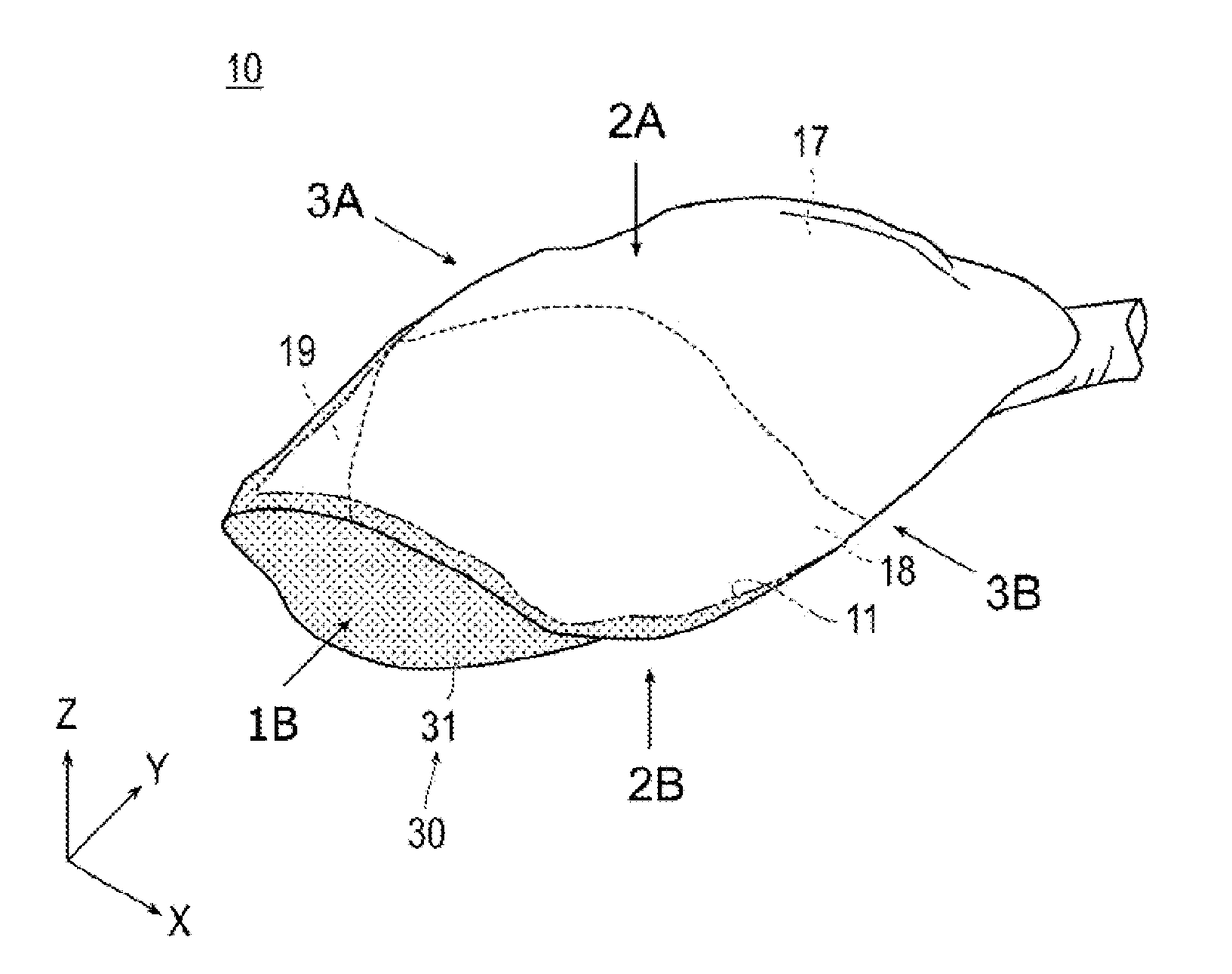
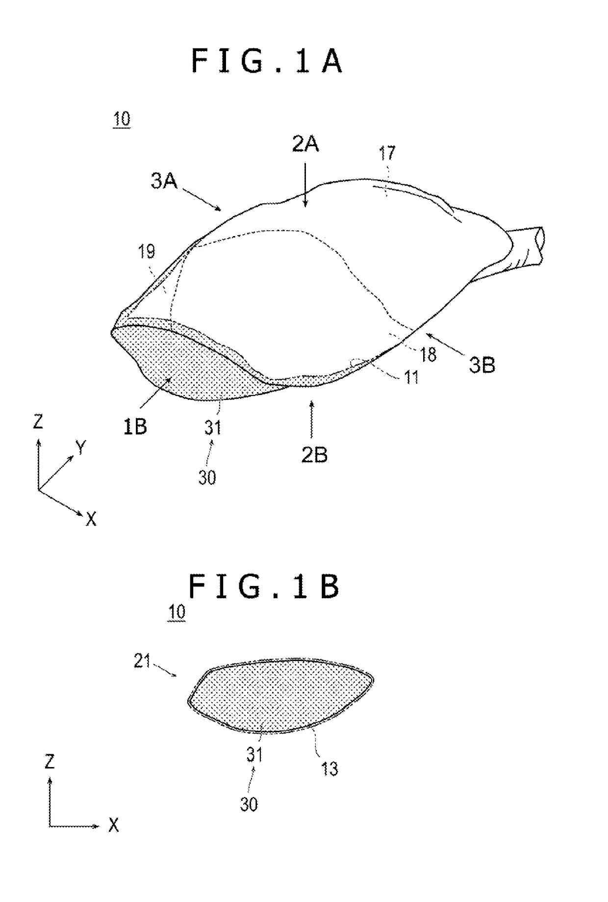
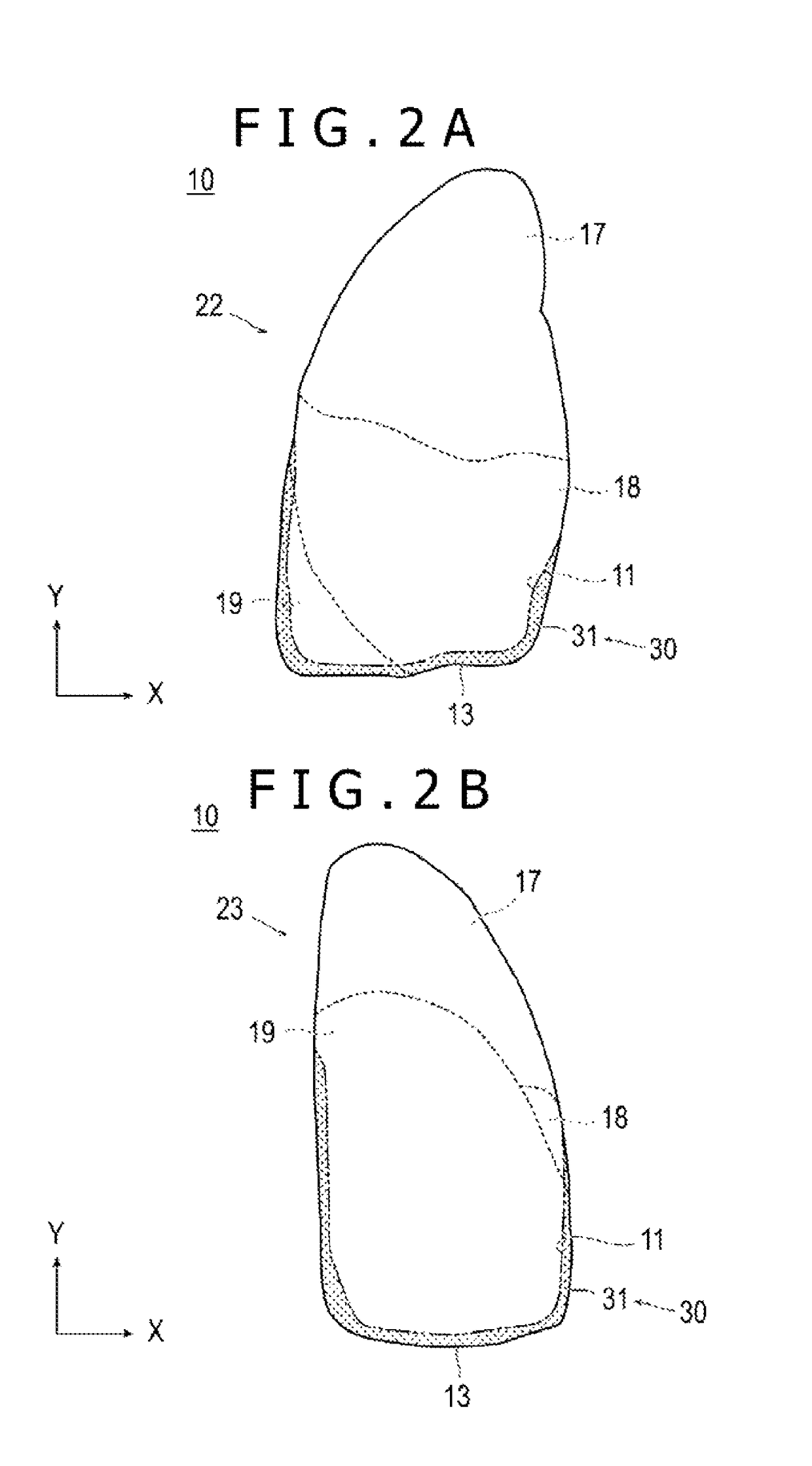
[0077]FIG. 9 illustrates schematically the result of a treatment performed on a lung 50 isolated from a pig, specifically, on a part similar to the target part 30 shown in FIGS. 1A to 3B. The present disclosure is not, however, limited to the mode described in this Example.

[0078]FIG. 9 shows the back side of the isolated lung 50 of the pig. In this Example, hot air at a high temperature was blown to the verge 11 of the surface of the pleura of the right lung 50 of the pig (to the part surrounded by two-dot chain line in the drawing), whereby the tissues and the lung parenchyma 15 in the surroundings of the verge 11 were cauterized. Incidentally, a mask member for prevention of cauterization by the hot air was disposed on other part than the verge 11. After the cauterization, the lung 50 is contained in a predetermined case, the inside of the case was deaerated, and fluid was fed into the inside of the l...


PUM


Abstract

A treatment method is provided which exhibits a reliable therapeutic effect on chronic obstructive pulmonary disease. In the treatment method, lung volume is reduced by restraining expansion and contraction of lung parenchyma present in the vicinity of a verge of a lung having an emphysema-suffering part, at least in one site of the verge.

Description

CROSS REFERENCE TO RELATED APPLICATION[0001]This application is based on Japanese Application No. 2013-169944, filed on Aug. 19, 2013, the contents of which are incorporated herein by reference.BACKGROUND[0002]The present disclosure relates to a method for the reduction of lung volume.[0003]Chronic obstructive pulmonary disease (COPD) means a wide variety of lung diseases that hinder normal breathing, and is a disease of lung obstruction caused by pulmonary emphysema or chronic bronchitis.[0004]Of these diseases, the pulmonary emphysema refers to a state of abnormal enlargement with destruction of tissues called pulmonary alveolar parenchyma inclusive of respiratory bronchioles, alveolar duct, pulmonary alveoli and alveolar sacs serving as sites of gas exchange. Although the pulmonary alveolar parenchyma in its normal state shrinks at the time of expiration, emphysema-suffering pulmonary alveolar parenchyma does not return into its original state if once enlarged by breathing. There...

Claims


Application Information

Patent Timeline
Patent Type & Authority Patents(United States)
IPC IPC(8): A61B18/04A61B18/02A61B18/00
CPCA61B18/04A61B18/02A61B2018/00595A61B2018/00541
Inventor HATA, SUGURUAKUTAGAWA, AYAKA
Owner TERUMO KK
Why Eureka
Eureka Blog
Learn More