


Quick Research
Generate reliable direction feasibility study reports for your R&D in just a few steps.

Technical Q&A
Discover and master advanced knowledge NOW. Basics, ideas, possibilities, all at once.

Find Solutions
As an expert in R&D theories, this can generate solutions to your technical problems instantly.

Evaluate Feasibility
Analyze your overall solution with one click, know your potential R&D risks in advance.

Monitor Landscape
Get weekly tech updates, stay abreast of the latest tech innovations and key insights.
Biological tissue motion trace method and image diagnosis device using the trace method
A technology of image diagnosis and tracking device, which is applied in the direction of diagnosis, image enhancement, image analysis, etc., and can solve the problems of loss of reliability and inability to become
- Summary
- Abstract
- Description
- Claims
- Application Information
AI Technical Summary
Problems solved by technology
Method used
Image

Examples
Embodiment 1
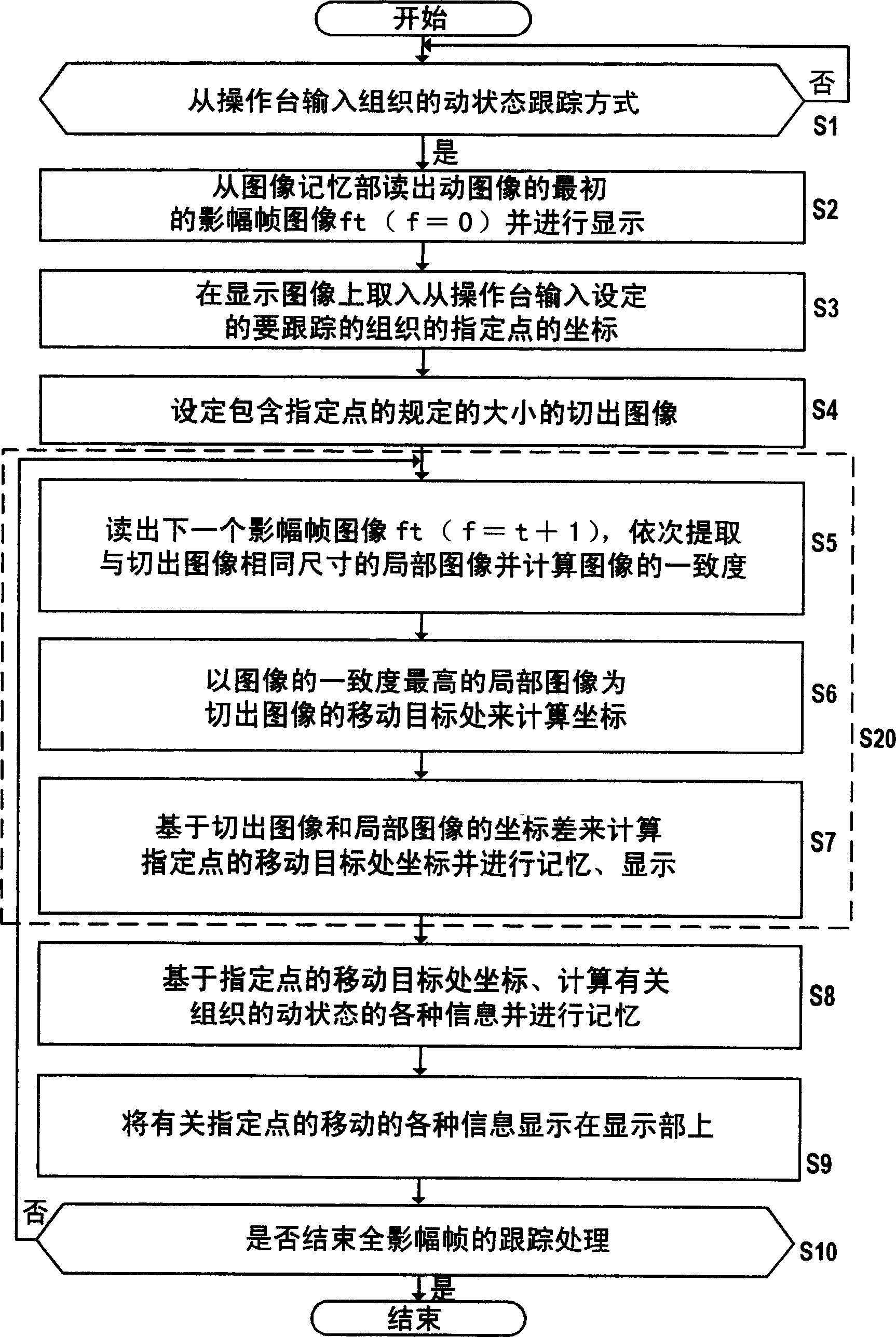
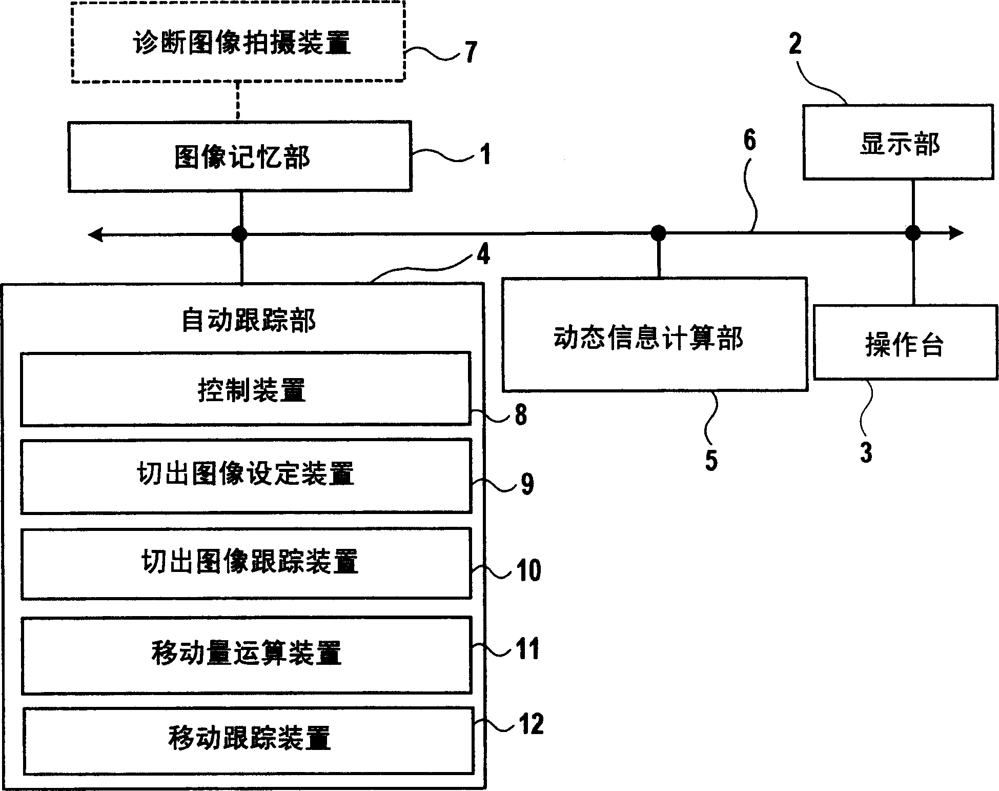
[0033] For the image diagnosis apparatus of one embodiment of the method for tracking the dynamic state of biological tissues of the present invention, use Figure 1 ~ Figure 4 Be explained. figure 1 Shows the procedure of the method for tracking the dynamic state of biological tissues of the present invention, figure 2 Is app figure 1 A block diagram of an image diagnosis apparatus constructed by a method for tracking the dynamic state of biological tissues. Such as figure 2 As shown, the image diagnosis apparatus includes the following parts: an image memory unit 1 that stores moving images of tomographic images of a subject, a display unit 2 that can display moving images, a console 3 for inputting instructions, and a tracking display on The automatic tracking unit 4 for the dynamic state of the living tissues of the moving image on the display unit 2, and the dynamic information calculating unit 5 for calculating various measurement information based on the tracking result...
Embodiment 2
[0050] figure 1 The example is to illustrate that whenever the tracking of a specified point of a frame image ends (S7), various information about the movement state of the tissue is calculated based on the movement of the specified point (S8). Example of information displayed on the display (S9). But the present invention is not limited to this, such as Picture 10 Shown, will figure 1 The step S10 of is arranged after the step S7, and the processing of the steps S8 and S9 can also be executed after the tracking of the designated points of all the frame images is completed.
[0051] Here, use the specific example of image tracking processing performed by the image correlation method Picture 11 Be explained. In the example of the figure, in order to simplify the description, the size of the cut-out image 25 is described as a rectangular 9-pixel area, and the search area 26 is also described as a rectangular 25-pixel area. That is, the cut-out image 25 shown in (a) is an exampl...
Embodiment 3
[0053] This embodiment can be applied to a moving image captured by an ultrasonic imaging method for tracking processing of a living body tissue. In particular, the RF signal corresponding to the moving image is memorized, and the position of the partial image with the highest degree of consistency of the image obtained by the image correlation method is corrected by using the RF signal to track the dynamic state of the biological tissue. The value changes smoothly.
[0054] in Picture 12 In particular, the case where the diagnostic image capturing device 7 is used as the ultrasonic diagnostic device 17 is shown. As the ultrasonic diagnostic apparatus 17, it transmits ultrasonic waves into the subject, receives the ultrasonic signals reflected by the tissues in the subject, processes the received signals, and displays the ultrasonic images in the subject based on the received signals to diagnose the subject’s disease And other diagnostic devices. The moving image given from the u...
PUM
 Abstract
Description
Claims
Application Information
 - R&D Engineer
- R&D Manager
- IP Professional
- Industry Leading Data Capabilities
- Powerful AI technology
- Patent DNA Extraction
Browse by: Latest US Patents, China's latest patents, Technical Efficacy Thesaurus, Application Domain, Technology Topic, Popular Technical Reports.
© 2024 PatSnap. All rights reserved.Legal|Privacy policy|Modern Slavery Act Transparency Statement|Sitemap|About US| Contact US: help@patsnap.com