Method of detecting 5'-methioadenosine phosphorylase defect type mammarian cell
A technology of polynucleotides and immune animals, applied in the direction of microorganism-based methods, botanical equipment and methods, biochemical equipment and methods, etc., can solve the problems of low yield, lack of detection, laborious and other problems
- Summary
- Abstract
- Description
- Claims
- Application Information
AI Technical Summary
Problems solved by technology
Method used
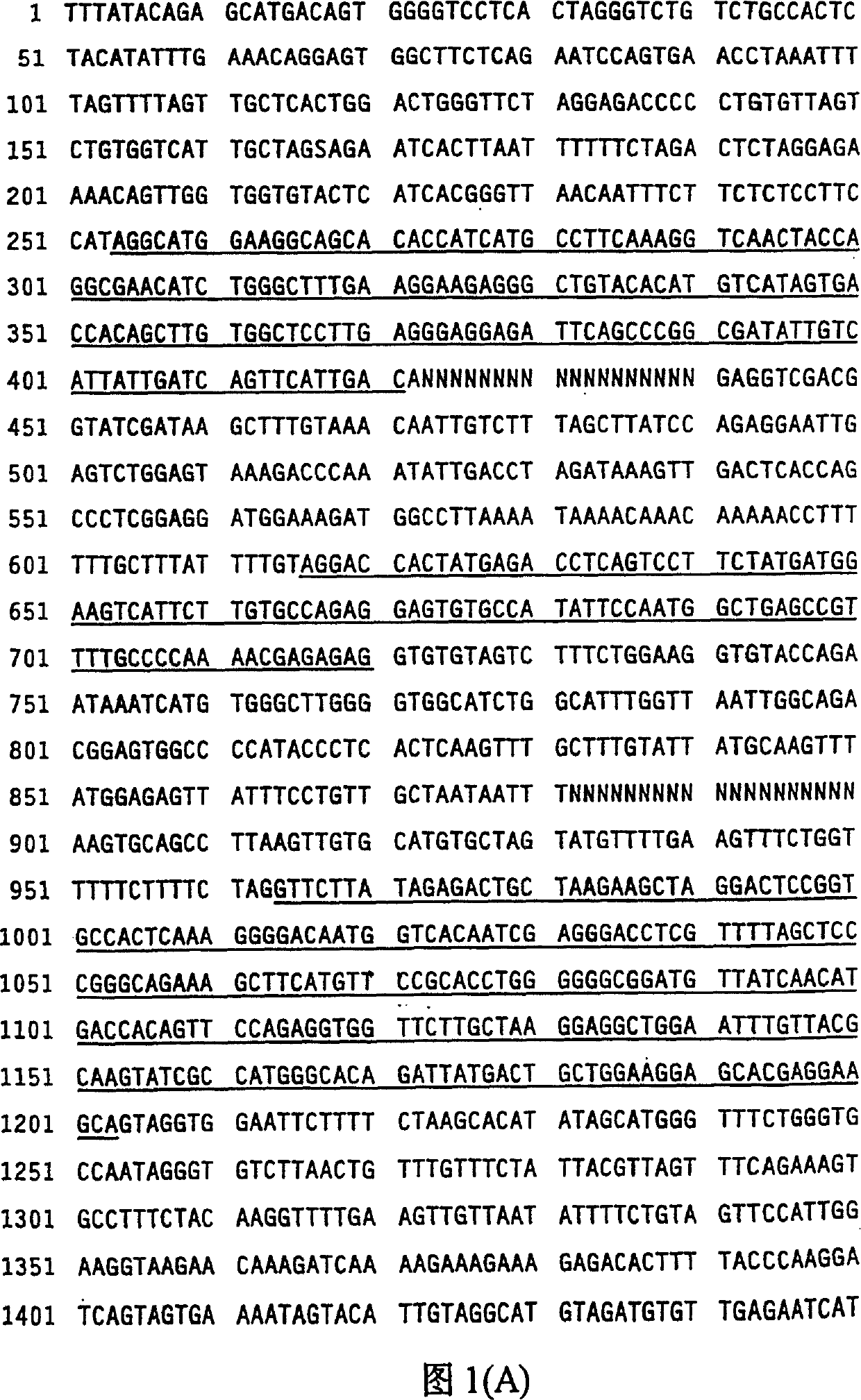
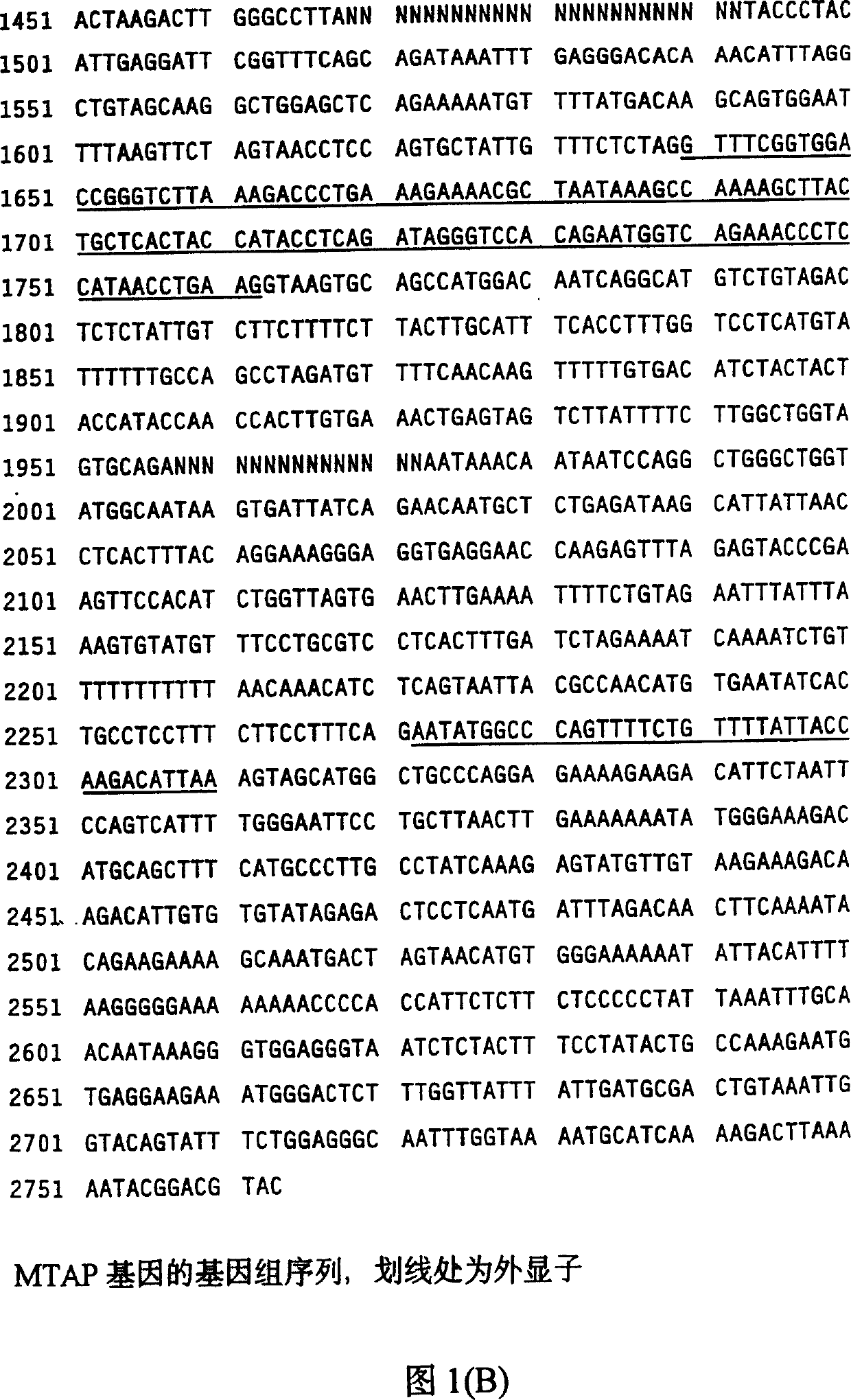
Image

Examples
Embodiment I
[0071] Detection of MTA enzyme catalytic activity in one sample
[0072] By detection from [methyl- 14 C] 5′-deoxy-5′-methylthioadenosine [methyl- 14 C] 5-Methylthioribose-1-phosphate can measure the phosphorolytic activity of MTA enzymes (Seidenfeld et al., Biochem. Biophys. Res. Commun. 95, 1861-1866, 1980). In a total volume of 200 μl, the standard reaction mixture contained 50 mM potassium phosphate buffer, pH 7.4, 0.5 mM [methyl- 14 C] 5'deoxy-5'-methylthioadenosine (2×10 5 CPM / mmol), 1 mM DTT and the indicated amounts of enzyme. After incubation at 37°C for 20 minutes, the reaction was terminated by adding 50 μl of 3M trichloroacetic acid, and 200 μl of the sample was added to a 0.6×2 cm “Dowex” 50-H equilibrated with water. * column. Will [methyl- 14 C] 5-Methylthioribose-1-phosphate was eluted directly into scintillation vials containing 2 ml of 0.1 M HCl.
Embodiment II
[0074] Purification of Native MTAase from Rat Liver
[0075] MTAse was isolated from rat liver using a modification of the method of Rangione et al. (J. Biol. Chem. 261, 12324-12329, 1986). 50 g of fresh rat liver was homogenized in Waring Blendor with 4 times the volume of 10 mM potassium phosphate buffer containing 1 mM DTT, pH 7.4 (buffer A). The homogenate was centrifuged (15,000 x g for 1 hour), and the resulting supernatant was subjected to ammonium sulfate fractionation. The pellet between 55 and 75% saturation was collected by centrifugation (15,000 xg for 20 minutes) and dissolved in a minimal volume of buffer A. Samples were then dialyzed overnight against three changes of 100 volumes of the same buffer.
[0076] Samples were clarified by centrifugation at 15,000 xg for 30 min and loaded onto a DEAE-Sephacyl column (1.5 x 18 cm; Pharmacia) pre-equilibrated with buffer A. After washing with 80 ml equilibration buffer, it was eluted with a linear ...
Embodiment III
[0079] Determination of Partial Amino Acid Sequence of Rat MTAzyme
[0080] The purified sample was lyophilized, dissolved in 50 μl carrier buffer (1% + sodium dialkyl sulfate (SDS), 10% glycerol, 0.1M DTT and 0.001% bromophenol blue) and loaded onto 0.5 mm thick 10% SDS poly Acrylamide gel (Bio Rad "MINIGEL" apparatus). After electrophoresis, according to Towbin et al. described (Proc.Natl.Acad.Sci.USA 76,4340-4345,1979) using transfer buffer (15mM Tris, 192mM glycine and 20% methanol, pH8.3) in a Bio-Rad The protein was electroblotted for 2 hours onto a nitrocellulose membrane (0.45 [mu]m pore size, Millipore) in a transfer system.
[0081] After transfer, proteins were reversibly stained with Ponceau S (Sigma) following a modified method described by Sailinovich and Montelaro (Anal. Biochem. 156, 341-347, 1987). The nitrocellulose membrane was then dipped for 60 seconds in a solution of 0.1% Ponsean S dye in 1% aqueous acetic acid. Excess dye was removed ...
PUM
 Abstract
Description
Claims
Application Information
 - R&D
- Intellectual Property
- Life Sciences
- Materials
- Tech Scout
- Unparalleled Data Quality
- Higher Quality Content
- 60% Fewer Hallucinations
Browse by: Latest US Patents, China's latest patents, Technical Efficacy Thesaurus, Application Domain, Technology Topic, Popular Technical Reports.
© 2025 PatSnap. All rights reserved.Legal|Privacy policy|Modern Slavery Act Transparency Statement|Sitemap|About US| Contact US: help@patsnap.com