Thyroid nodule ultrasound image segmentation method, device and system
A technology of thyroid nodules and ultrasound images, applied in image analysis, image data processing, neural learning methods, etc., can solve the problem of low accuracy of thyroid nodule image segmentation, and achieve the effect of improving segmentation accuracy and accuracy
- Summary
- Abstract
- Description
- Claims
- Application Information
AI Technical Summary
Problems solved by technology
Method used
Image


Examples
specific Embodiment 1
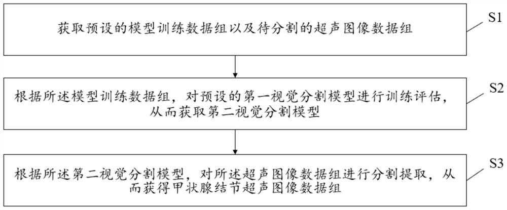
[0027] The embodiment of the present invention firstly describes a method for segmenting an ultrasound image of a thyroid nodule. figure 1 A flowchart of an embodiment of a method for segmenting an ultrasound image of a thyroid nodule according to the present invention is shown.
[0028] like figure 1 As shown, the method includes the following steps:
[0029] S1: Obtain a preset model training data set and an ultrasound image data set to be segmented.
[0030] In order to improve the segmentation accuracy of the thyroid nodule region in the ultrasound image, the embodiment of the present invention proposes to improve the existing semantic segmentation network Unet, and introduce a preset hierarchical cascade multi-head self-attention module (H -MHSA module) and modify the original pooling layer downsampling to a dual-branch downsampling method, and train the improved model, and then use the trained model to treat the segmented ultrasound image (for ease of description and u...
specific Embodiment 2
[0043] Furthermore, the embodiment of the present invention also describes a method for segmenting an ultrasound image of a thyroid nodule. figure 2 A flowchart showing another embodiment of a method for segmenting an ultrasound image of a thyroid nodule according to the present invention.
[0044] like figure 2 As shown, the method includes the following steps:
[0045] A1: Replace the downsampling method of the preset semantic segmentation network Unet with the two-branch downsampling method to obtain the first improved model.
[0046] Among them, the two-branch downsampling method means that one branch is downsampled through a typical 3x3 convolution with a step size of 2, and the other branch is downsampled through maximum pooling and combined with 1x1 convolution for channel transformation. The two branches The information of Add is fused by element-by-element summation, which can retain more context information. In one embodiment, the preset RelU activation function...
specific Embodiment 3
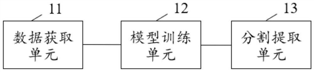
[0069] In addition to the above method, the embodiment of the present invention also describes a device for segmenting ultrasound images of thyroid nodules. image 3 A structural diagram of an embodiment of a device for segmenting an ultrasound image of a thyroid nodule according to the present invention is shown.
[0070] As shown in the figure, the segmentation device includes a data acquisition unit 11 , a model training unit 12 and a segmentation extraction unit 13 .
[0071] The data acquiring unit 11 is used to acquire preset model training data sets and ultrasound image data sets to be segmented.
[0072] The model training unit 12 is configured to train and evaluate the preset first visual segmentation model according to the model training data set, so as to obtain the second visual segmentation model. The first visual segmentation model is obtained by introducing a preset hierarchical cascaded multi-head self-attention module and modifying the original pooling layer ...
PUM
 Abstract
Description
Claims
Application Information
 - R&D
- Intellectual Property
- Life Sciences
- Materials
- Tech Scout
- Unparalleled Data Quality
- Higher Quality Content
- 60% Fewer Hallucinations
Browse by: Latest US Patents, China's latest patents, Technical Efficacy Thesaurus, Application Domain, Technology Topic, Popular Technical Reports.
© 2025 PatSnap. All rights reserved.Legal|Privacy policy|Modern Slavery Act Transparency Statement|Sitemap|About US| Contact US: help@patsnap.com