Ocular Surface Fluid Collection and Detection Device
A detection device and ocular surface technology, which is applied in the field of medical devices, can solve the problems of judging the patient's eye disease, the discomfort of the collected person, and the impact of the detection result, so as to avoid the influence of repeated freezing and thawing, short detection time, simple and convenient operation Effect
- Summary
- Abstract
- Description
- Claims
- Application Information
AI Technical Summary
Problems solved by technology
Method used
Image
Examples
Embodiment 1
[0083] This embodiment provides an ocular surface fluid collection and detection device, including a collection part and a detection part;
[0084] Such as figure 1 with figure 2 As shown, the collection part includes a casing, a groove 1 arranged on the outer surface of the casing for collecting ocular surface fluid, and a suction hole 2 arranged at the bottom of the groove.
[0085] The groove 1 is arranged at one end of the casing, and the groove 1 forms an opening with the edge of the casing.
[0086] The groove 1 is also provided with a drainage groove 3 , one end of the drainage groove 3 is connected to the edge of the sample suction hole 2 , and the other end extends toward the opening of the groove 1 .
[0087] The outermost edge of the groove is also provided with an anti-overflow strip 4 .
[0088] Such as image 3 with Figure 4 As shown, the housing includes a card cover and a card bottom:
[0089] Both the inner surface of the card cover and the inner surfa...
Embodiment 2
[0104] This embodiment provides the method for detecting the specific IgE antibody in the ocular surface fluid sample to be tested by using the colloidal gold qualitative method in embodiment 1:
[0105] 1. Reaction principle
[0106] Anti-IgE monoclonal antibody No. 1 (T line) and goat anti-chicken polyclonal antibody (C line) are pre-immobilized on the nitrocellulose membrane of the immunochromatographic detection card, and anti-IgE monoclonal antibody 2 pre-coated with colloidal gold on the gold standard pad No. and Chicken IgY. After the collected sample of the ocular surface fluid to be tested enters the suction hole 2, the sample of the ocular surface fluid to be tested flows through the sample pad, the gold standard pad and the nitrocellulose membrane according to capillary action. When the sample contains IgE, the target object first forms a reaction complex with the colloidal gold-labeled anti-IgE monoclonal antibody No. 2, and forms a double-antibody sandwich comple...
Embodiment 3
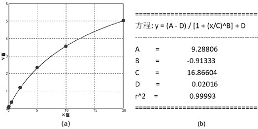
[0125] This embodiment provides the method of detecting the specific IgE antibody in the ocular surface fluid sample to be tested by using the fluorescence quantitative method in embodiment 1:
[0126] 1. Reaction principle
[0127] Anti-IgE monoclonal antibody No. 1 (line T) and goat anti-chicken polyclonal antibody (line C) are pre-fixed on the nitrocellulose membrane, anti-IgE monoclonal antibody No. 2 and chicken polyclonal antibody pre-coated with fluorescent microspheres are pre-coated on the fluorescent pad IgY. After the collected ocular surface fluid sample to be tested enters the suction hole, the ocular surface fluid sample to be tested flows through the sample pad, the fluorescent pad and the nitrocellulose membrane according to capillary action. When the sample contains IgE, the target object first forms a reaction complex with the fluorescently labeled anti-IgE monoclonal antibody No. 2, and forms a double antibody sandwich complex with it when passing through t...
PUM
 Abstract
Description
Claims
Application Information
 - R&D
- Intellectual Property
- Life Sciences
- Materials
- Tech Scout
- Unparalleled Data Quality
- Higher Quality Content
- 60% Fewer Hallucinations
Browse by: Latest US Patents, China's latest patents, Technical Efficacy Thesaurus, Application Domain, Technology Topic, Popular Technical Reports.
© 2025 PatSnap. All rights reserved.Legal|Privacy policy|Modern Slavery Act Transparency Statement|Sitemap|About US| Contact US: help@patsnap.com


