Heterotopic ossification CT image segmentation method and device, and heterotopic ossification CT image segmentation three-dimensional reconstruction method and device

A technology of CT images and abnormal images, which is applied in the field of medical image processing, can solve the problems that limit the efficiency of viewing films, such as the progress of patients with heterotopic ossification, and achieve the effect of reducing the difficulty of acquisition and improving the speed

Pending Publication Date: 2021-07-09
XIANGYA HOSPITAL CENT SOUTH UNIV
View PDF7 Cites 2 Cited by
  • Summary
  • Abstract
  • Description
  • Claims
  • Application Information

AI Technical Summary

Problems solved by technology

However, because the images of heterotopic ossification are similar to those of normal bones, it requires extensive experience in clinical imaging to identify the site of heterotopic ossification from CT scan images, and it is difficult for young or inexperienced doctors to accurately find it. It greatly limits the efficiency of doctors in seeing films and predicting the progress of patients with heterotopic ossification

Method used


Image

  • Heterotopic ossification CT image segmentation method and device, and heterotopic ossification CT image segmentation three-dimensional reconstruction method and device
  • Heterotopic ossification CT image segmentation method and device, and heterotopic ossification CT image segmentation three-dimensional reconstruction method and device
  • Heterotopic ossification CT image segmentation method and device, and heterotopic ossification CT image segmentation three-dimensional reconstruction method and device

Examples


Embodiment Construction

[0052] In order to make the purpose, technical solution and advantages of the present invention clearer, the technical solution of the present invention will be described in detail below. Apparently, the described embodiments are only some of the embodiments of the present invention, but not all of them. Based on the embodiments of the present invention, all other implementations obtained by persons of ordinary skill in the art without making creative efforts fall within the protection scope of the present invention.

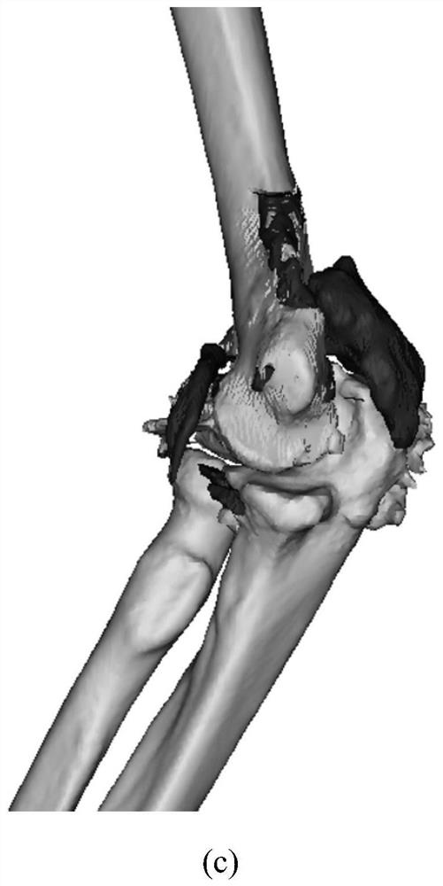
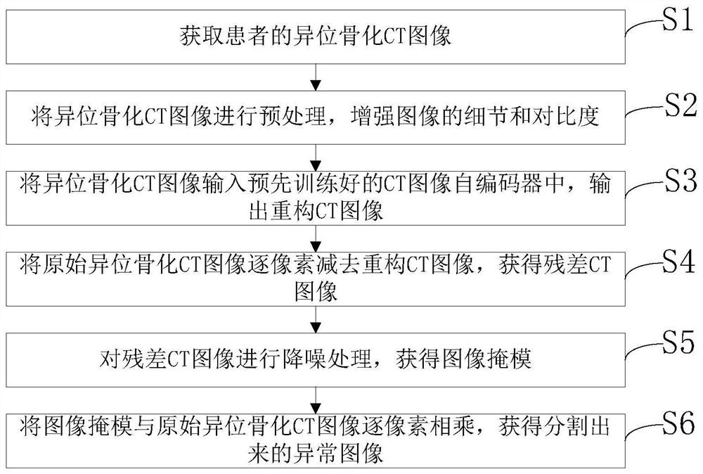
[0053] Such as figure 2 and image 3 As shown, the embodiment of the present invention provides a heterotopic ossification CT image segmentation method. Before performing segmentation processing, a CT image autoencoder needs to be trained. The input of the CT image autoencoder is a CT image. The output is a reconstructed image. Specifically, the CT image self-encoder is trained by the following method:

[0054] A1: Obtain multiple CT images of normal people t...


PUM


Abstract

The invention discloses a heterotopic ossification CT image segmentation method and device, and a heterotopic ossification CT image segmentation three-dimensional reconstruction method and device. The segmentation method comprises the following steps: acquiring a heterotopic ossification CT image of a patient; inputting the heterotopic ossification CT image into a pre-trained CT image auto-encoder, and outputting a reconstructed CT image; subtracting the reconstructed CT image from the original heterotopic ossification CT image pixel by pixel to obtain a residual CT image; obtaining an abnormal region through noise reduction processing; transforming the abnormal region into an image mask consistent with the original heterotopic ossification CT image in size; and multiplying the image mask by the original heterotopic ossification CT image pixel by pixel to obtain a segmented abnormal image. According to the scheme, the heterotopic ossification part can be automatically segmented from the CT image, three-dimensional reconstruction of the heterotopic ossification stereoscopic view can be facilitated, the film viewing speed of doctors can be greatly improved, and low-age doctors or inexperienced people can be helped to accurately find the heterotopic ossification part.

Description

technical field [0001] The invention relates to the technical field of medical image processing, in particular to a heterotopic ossification CT image segmentation method, a three-dimensional reconstruction method, and a device. Background technique [0002] To check a patient for the presence of heterotopic ossification, a CT scan is usually done. Such as figure 1 (a) shows the cross section of CT scan of heterotopic ossification, and the framed part is the position of heterotopic ossification, figure 1 (b) Sagittal CT scan of heterotopic ossification, where the framed part is the position of heterotopic ossification, figure 1 (c) CT scan for heterotopic ossification Figure three Stereoscopic view obtained after three-dimensional reconstruction, where the dark part is the heterotopic ossification site after three-dimensional reconstruction. However, because the images of heterotopic ossification are similar to those of normal bones, it requires extensive experience in c...

Claims


Application Information

Patent Timeline
IPC IPC(8): G06T7/00G06T7/11G06T5/00G06T17/00G06N3/04G06N3/08
CPCG06T7/0012G06T7/11G06T17/00G06N3/084G06T2207/10081G06T2207/20081G06T2207/20084G06T2207/30008G06N3/045G06T5/70
Inventor 成亮江梦林涨源朱勇
Owner XIANGYA HOSPITAL CENT SOUTH UNIV
Why Eureka
Try Eureka