Elastic imaging method and device, electronic equipment and storage medium

An elastography and viscoelasticity technology, applied in medical science, diagnosis, application, etc., can solve the problems of poor imaging effect and low precision, achieve rich frequency components, good response effect, and improve imaging effect and imaging accuracy.

Pending Publication Date: 2021-07-09
WUXI HISKY MEDICAL TECH +1
View PDF3 Cites 1 Cited by
  • Summary
  • Abstract
  • Description
  • Claims
  • Application Information

AI Technical Summary

Problems solved by technology

[0005] The invention provides an elastography method, device, electronic equipment and storage medium to solve the problems of poor imaging effect and low precision in the existing elastography

Method used


Image

  • Elastic imaging method and device, electronic equipment and storage medium
  • Elastic imaging method and device, electronic equipment and storage medium
  • Elastic imaging method and device, electronic equipment and storage medium

Examples


Embodiment Construction

[0045] Reference will now be made in detail to the exemplary embodiments, examples of which are illustrated in the accompanying drawings. When the following description refers to the accompanying drawings, the same numerals in different drawings refer to the same or similar elements unless otherwise indicated. The implementations described in the following exemplary examples do not represent all implementations consistent with the present disclosure. Rather, they are merely examples of apparatuses and methods consistent with aspects of the present disclosure as recited in the appended claims.

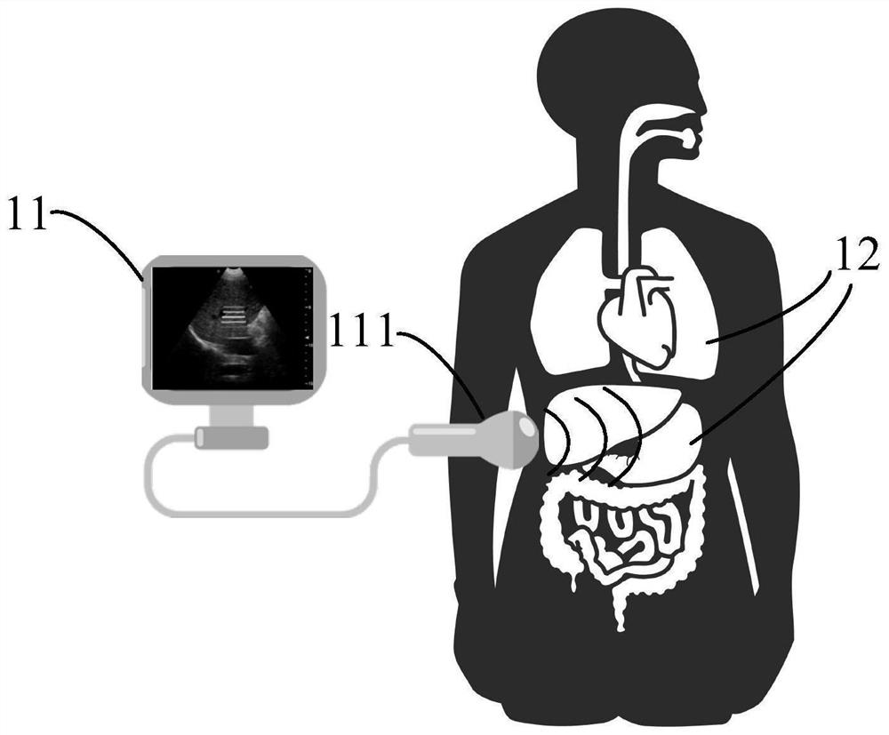
[0046] The application scenarios of the embodiments of the present invention are explained below:

[0047] figure 1 An application scene diagram of the elastic imaging method provided by the embodiment of the present invention, such as figure 1 As shown, the elastography method provided by the embodiment of the present invention is applied to an electronic device, specifically, for e...


PUM


Abstract

The invention provides an elastic imaging method and device, electronic equipment and a storage medium. The method comprises the steps: obtaining preset vibration excitation parameters which comprise at least two different vibration frequencies; sequentially applying vibration excitation with different vibration frequencies to the to-be-detected tissue, and performing ultrasonic detection; according to the detected ultrasonic echo signals corresponding to different vibration frequencies, viscoelasticity information of the to-be-detected tissue is calculated, due to the fact that the interior of the biological tissue is not of a uniform tissue structure and has difference in structural attribute, forced vibration responses generated after different tissue structures are excited by different frequencies are different, and the viscoelasticity information of the to-be-detected tissue is obtained. During viscoelasticity analysis, mechanical vibration with different frequencies is utilized to excite tissue structures in biological tissues, so that frequency parts are richer, different tissue structures can generate better response effects, and the imaging effect and the imaging precision of elastography are further improved.

Description

technical field [0001] The invention relates to the technical field of medical equipment, in particular to an elastic imaging method, device, electronic equipment and storage medium. Background technique [0002] Ultrasound imaging technology is widely used in clinical diagnosis because of its advantages of real-time, cheap, non-invasive and non-ionizing radiation. By obtaining the physiological structure information of biological tissues, it can realize the detection of lesions in biological tissues. However, in the early lesions of biological tissues, the traditional B-ultrasound and other ultrasonic imaging methods are not sensitive to the early lesions of biological tissues because their structure does not change significantly. [0003] Elastic imaging technology is a new imaging technology that has emerged in recent years. It excites the vibration of biological tissue through shear waves and other methods, and uses ultrasonic signals to track the deformation information...

Claims


Application Information

Patent Timeline
Patent Type & Authority Applications(China)
IPC IPC(8): A61B8/00
CPCA61B8/485A61B8/5215A61B8/5261
Inventor 何琼邵金华孙锦
Owner WUXI HISKY MEDICAL TECH
Why Eureka
Try Eureka