Method for improving infection efficiency of adeno-associated virus to infect cells
A technology for infecting cells and cells, which is applied in the field of biomedicine and can solve problems such as restricting the application of AAV and weak capabilities
- Summary
- Abstract
- Description
- Claims
- Application Information
AI Technical Summary
Problems solved by technology
Method used
Image
Examples
Embodiment approach
[0131] CLAIMS 1. A method for increasing the infection efficiency of an adeno-associated virus (AAV)-infected cell, comprising the step of: contacting said cell to be infected with a DNA replication inhibitor.
[0132] 2. The method of embodiment 1, wherein the contacting comprises contacting in vitro.
[0133] 3. The method of any one of embodiments 1-2, wherein the contacting time is from about 0.5 to about 4 hours.
[0134] 4. The method of any one of embodiments 1-3, wherein the temperature of the contacting is from about 30°C to about 40°C.
[0135] 5. The method according to any one of embodiments 1-4, wherein said cells to be infected are contacted with a DNA replication inhibitor in a cell culture medium.
[0136] 6. The method according to embodiment 5, wherein the cell culture medium comprises DMEM medium, HAM'S F12K medium and / or FBS medium.
[0137] 7. The method according to any one of embodiments 1-6, wherein the cells comprise: HEK293 cells, 293T cells and / or ...
Embodiment 1
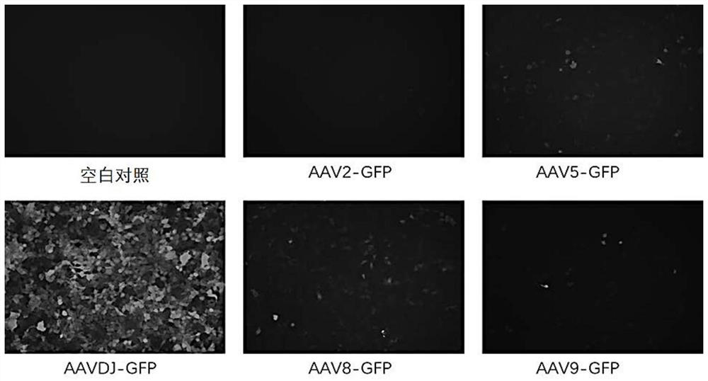
[0171] Example 1 Different serotypes of AAV-GFP have different infectivity and expression intensity in HEK293
[0172] HEK293 cells (ATCC, CRL-1573) were digested 24 hours in advance, and 12-well cell culture dishes (Corning, 3513) were incubated with 100 μg / ml poly-lysine (Beyond, C0313)-DPBS (Gibco, C14190500BT) solution in advance After 15 minutes, the cells were blotted dry, and the cells were counted at 1.5e5 per well. CO 2 Incubate at 37°C for 24 hours in an incubator.
[0173] After 24 hours, each well was infected with AAV2-GFP, AAV5-GFP, AAV8-GFP, AAV9-GFP and AAVDJ-GFP at MOI=3E5 respectively (wherein each AAV was purchased from Shandong Weizhen Biotechnology Co., Ltd. The described steps allow AAV to express GFP (the accession number of the GFP gene in GenBank is ID: 114976950)). Fresh DMEM-10% FBS (Gibco, 10199-141) medium was replaced every 24 hours after infection. After 2 days of infection, a fluorescent microscope (Life, AMF4305) was used to take pictures a...
Embodiment 2
[0175] Example 2 Different serotypes of AAV-GFP have different infectivity and expression intensity in CHO cells
[0176] Digest CHO-K1 cells (Wuhan Punuo, CL-0062) 24 hours in advance, and 12-well cell culture dishes (Corning, 3513) were pre-digested with 100 μg / ml poly-lysine (Gibco , C14190500BT) solution was incubated for 15min, blotted dry and plated cells, counted 1.5e5 per well. CO 2 Incubate at 37°C for 24 hours in an incubator.
[0177] After 24 hours, each well was infected with AAV2-GFP, AAV5-GFP, AAV8-GFP, AAV9-GFP, and AAVDJ-GFP at MOI=3E5, respectively. Fresh Ham's F12K (Wuhan Punuosai, PM150910)-10% FBS medium was replaced every 24 hours after infection. After 3 days of infection, photographs were taken with a fluorescent microscope, and the fluorescent expression level of GFP was detected with a microplate reader (Biotek, H1M).
[0178] The expression levels of GFP fluorescence after different serotypes of AAV-GFP infected CHO-K1 were as follows: Figure 3...
PUM
 Abstract
Description
Claims
Application Information
 - R&D Engineer
- R&D Manager
- IP Professional
- Industry Leading Data Capabilities
- Powerful AI technology
- Patent DNA Extraction
Browse by: Latest US Patents, China's latest patents, Technical Efficacy Thesaurus, Application Domain, Technology Topic, Popular Technical Reports.
© 2024 PatSnap. All rights reserved.Legal|Privacy policy|Modern Slavery Act Transparency Statement|Sitemap|About US| Contact US: help@patsnap.com


