
SA-Net-based MRI medical image segmentation method

A medical image and image technology, applied in the field of MRI medical image processing, can solve the problem of not being able to make full use of full-scale information

Pending Publication Date: 2021-06-11
山西三友和智慧信息技术股份有限公司
View PDF0 Cites 3 Cited by
  • Summary
  • Abstract
  • Description
  • Claims
  • Application Information

AI Technical Summary

Problems solved by technology

[0004] Aiming at the technical problem that the above-mentioned scale-based feature fusion cannot make full use of full-scale information, the present invention provides an MRI medical image segmentation method based on SA-Net with full utilization, high efficiency and strong reliability

Method used


Image

  • SA-Net-based MRI medical image segmentation method

Examples


Embodiment Construction

[0023] The following will clearly and completely describe the technical solutions in the embodiments of the present invention with reference to the accompanying drawings in the embodiments of the present invention. Obviously, the described embodiments are only some, not all, embodiments of the present invention. Based on the embodiments of the present invention, all other embodiments obtained by persons of ordinary skill in the art without making creative efforts belong to the protection scope of the present invention.

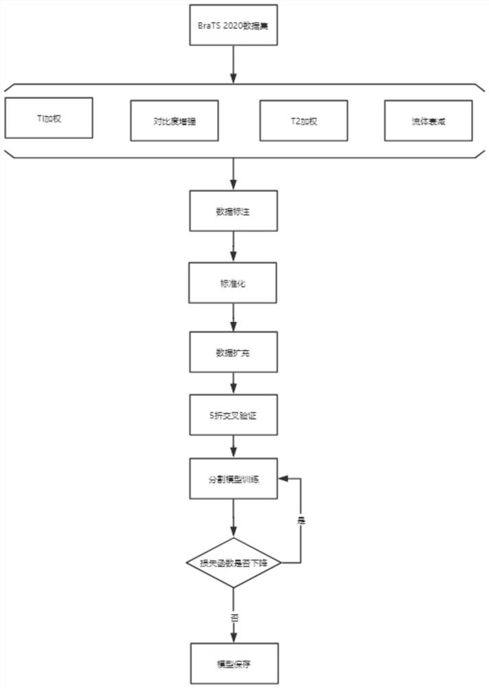
[0024] An MRI medical image segmentation method based on SA-Net, such as figure 1 shown, including the following steps:

[0025] S1. BraTS 2020 data collection: By collecting the dataset provided by BraTS 2020, which collected MRI scans from 19 institutions with different protocols, magnetic field strengths, and manufacturers. Each patient underwent native T1-weighted imaging, contrast-enhanced imaging, T2-weighted imaging, and fluid-attenuated imaging. The ...


PUM


Abstract

The invention belongs to the field of MRI medical image processing, and particularly relates to an SA-Net-based MRI medical image segmentation method, which comprises the following steps: BraTS 2020 data acquisition: acquiring a primary T1 weighted imaging data set, a contrast enhancement imaging data set, a T2 weighted imaging data set and a fluid attenuation imaging data set provided by BraTS 2020; data annotation: manually annotating the data set according to the same annotation protocol; data preprocessing: preprocessing the MRI image; training a segmentation model: segmenting the MRI medical image by using the variation of the U-Net model; adjusting model parameters through a loss function, obtaining an optimal model, and finishing the construction process of the segmentation model; and when the loss function of the model is not reduced any more, storing the model. According to the invention, performance evaluation is carried out on the model through five-fold cross validation, and full-scale information can be fully utilized to carry out MRI medical image segmentation. The invention is used for segmentation of the MRI medical image.

Description

technical field [0001] The invention belongs to the field of MRI medical image processing, in particular to a method for segmenting MRI medical images based on SA-Net. Background technique [0002] At present, the automatic segmentation of medical images is used to extract quantitative imaging biomarkers for accurate lesion site detection, which is a key step in diagnosis, prognosis, treatment planning and evaluation, and it is also the most challenging step. Multiparametric magnetic resonance imaging (mpMRI), used as a primary imaging modality for cancer treatment, provides a variety of different tissue properties. However, correctly interpreting mpMRI images is a challenging task, not only because mpMRI sequences generate a large amount of 3D or 4D image data, but also because of the inherent heterogeneity of MRI medical images. Therefore, there is an increasing demand for computerized analysis, which can assist clinicians to better interpret the lesion location in mpMRI ...

Claims


Application Information

Patent Timeline
IPC IPC(8): G06T7/10
CPCG06T7/10G06T2207/10088G06T2207/20081G06T2207/20084G06T2207/30096
Inventor 潘晓光张海轩刘剑超宋晓晨王小华
Owner 山西三友和智慧信息技术股份有限公司
Learn More