
Simple in-vitro liver perfusion device for digesting and separating liver cells

A simple technology for liver cells, which is applied in the field of simple extracorporeal liver perfusion devices, can solve the problems of large damage to liver cells and difficulty in extraction, and achieve the effect of accelerating separation efficiency and facilitating separation

Active Publication Date: 2021-02-26
GUILIN MEDICAL UNIVERSITY
View PDF16 Cites 1 Cited by
  • Summary
  • Abstract
  • Description
  • Claims
  • Application Information

AI Technical Summary

Problems solved by technology

[0004] The purpose of the present invention is to provide a simple extracorporeal liver perfusion device for digesting and separating liver cells, so as to solve the problems of large damage to liver cells and difficulty in extraction proposed in the above-mentioned background technology

Method used


Image

  • Simple in-vitro liver perfusion device for digesting and separating liver cells
  • Simple in-vitro liver perfusion device for digesting and separating liver cells
  • Simple in-vitro liver perfusion device for digesting and separating liver cells

Examples


Embodiment 1

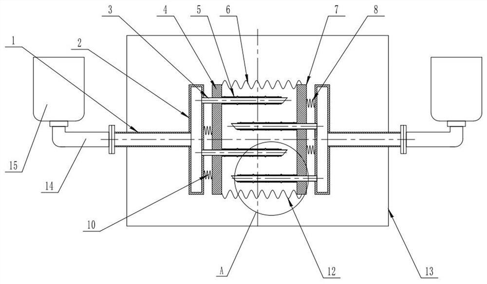
[0029]In order to clamp the liver tissue and quickly separate the liver cells from the tissue fluid, the present invention proposes a first embodiment, please refer tofigure 1 versusfigure 2 , The present invention provides a technical solution: a simple extracorporeal liver perfusion device for digestion and separation of hepatocytes, including a liver tissue separation sterile box 13 and a fixing mechanism, the fixing mechanism is installed in the liver tissue separation sterile box 13, and the fixing mechanism includes There are two extrusion units with the same structure on the left and right. The extrusion units are two parallel slides, the first slide 4 and the second slide 7, respectively. After the perfusion fluid is introduced into the liver tissue, the two slides are placed on the elastic member. Under the action of, it is relatively close together to squeeze the liver tissue in the squeezing space, and accelerate the separation efficiency of liver cells in the liver tissu...

Embodiment 2

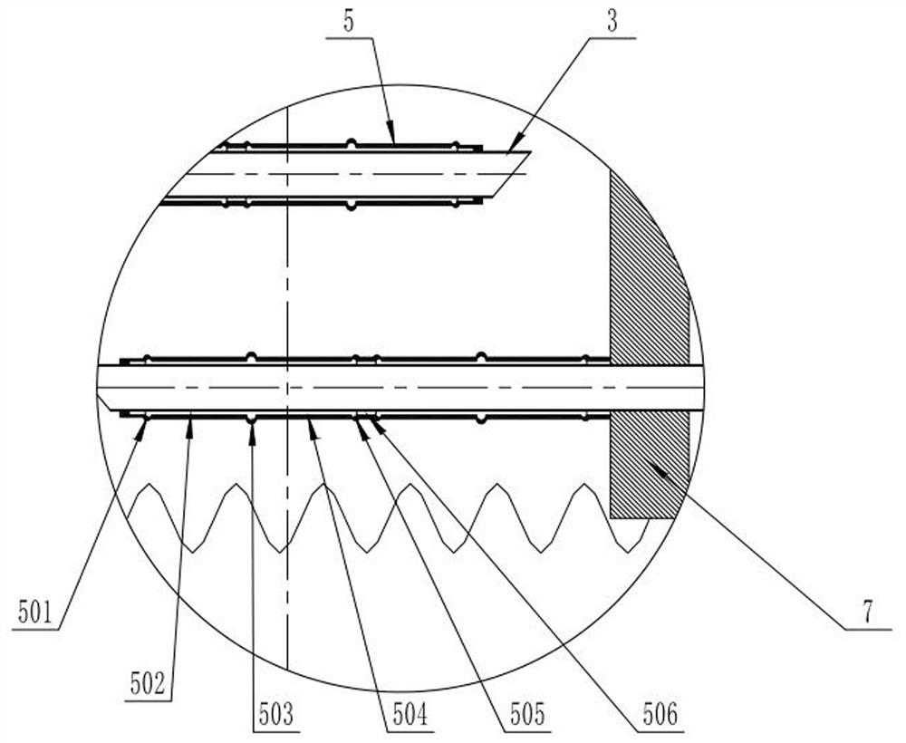
[0037]In order to assist the separation of liver tissue and make the separation more thorough, the present invention proposes another embodiment. A sleeve 5 that can be expanded to the outside is sleeved on the outside of the puncture needle 3, and one end of the sleeve 5 is fixed to the first sliding plate 4 The other end is fixedly connected to the end of the needle of the puncture needle 3. The sleeve 5 is provided with a plurality of expansion units and a plurality of sliding rings 506 sleeved on the outer wall of the puncture needle 3, and the adjacent expansion units pass through A slip ring 506 is connected. Each expansion unit includes four sets of supports. The four sets of supports are arranged in an annular distribution around the axis of the puncture needle 3. The slip ring 506 is slidingly sleeved on the needle tube body of the puncture needle 3. The blocking action of the first sliding plate 4 and the second sliding plate 7 expands the four groups of support bodies to ...


PUM


Abstract

The invention discloses a simple in-vitro liver perfusion device for digesting and separating liver cells. The simple in-vitro liver perfusion device comprises a liver tissue separation sterile box and a fixing mechanism, wherein the fixing mechanism is arranged in the liver tissue separation sterile box, the fixing mechanism comprises a left extrusion unit and a right extrusion unit with the samestructure, the liver tissue is placed between the two extrusion units, a puncture needle and a sleeve are inserted into the liver tissue, perfusate flows into the liver tissue from the puncture needle, the liver tissue is clamped through opposite movement of two extrusion units, meanwhile, multiple sets of expansion units are arranged on the sleeve outside the puncture needle, the expansion unitsare formed by movably connecting multiple pairs of supporting bodies with sliding rings capable of sliding through hinge points, when the two extrusion units get close to each other, the expansion units can automatically expand outwards under the action of thrust so as to clamp and fix the liver tissue till the liver tissue is completely separated, the separation time is shortened, and thereforethe liver tissue perfusion operation efficiency is further improved.

Description

Technical field[0001]The invention belongs to the related technical field of separating liver cells from liver tissue, and specifically relates to a simple extracorporeal liver perfusion device for digesting and separating liver cells.Background technique[0002]Hepatocytes are the main functional cells of the liver, mainly composed of deoxyribonucleic acid and histones. Hepatocytes are polygonal, with a diameter of about 20-30 / plus (micron), and 6-8 faces. The size of the liver cells varies under different physiological conditions. For example, the volume of liver cells becomes larger when starving, and the surface of each liver cell can be divided. There are three types: sinusoid surface, hepatocyte surface and bile duct surface.[0003]The existing simple extracorporeal liver perfusion device technology for digesting and separating hepatocytes has the following problems: the traditional simple extracorporeal liver perfusion device for digesting and separating hepatocytes requires sci...

Claims


Application Information

Patent Timeline
Patent Type & Authority Applications(China)
IPC IPC(8): C12M1/00
CPCC12M47/04C12M29/10C12M23/52C12M23/48
Inventor 邢雪琨李红男张佳阳
Owner GUILIN MEDICAL UNIVERSITY
Learn More