Renal pelvis pressure monitoring balloon perfusion catheter

A technology of pressure monitoring and balloons, which is applied in the direction of balloon catheters, catheters, diagnostic recording/measurement, etc., and can solve problems such as low cooling efficiency, no perfusion catheter, ice water leakage, etc.

Pending Publication Date: 2021-02-12
FUJIAN PROVINCIAL HOSPITAL
View PDF0 Cites 0 Cited by
  • Summary
  • Abstract
  • Description
  • Claims
  • Application Information

AI Technical Summary

Problems solved by technology

However, when the ordinary ureteral catheter is used for retrograde perfusion, there will still be a problem of rapid leakage of ice water, resulting in low cooling efficiency
[0004] There is currently no dedicated commercial perfusion catheter for renal pelvic ureter perfusion chemotherapy or ice water perfusion for renal hypothermia protection

Method used


Image

  • Renal pelvis pressure monitoring balloon perfusion catheter
  • Renal pelvis pressure monitoring balloon perfusion catheter
  • Renal pelvis pressure monitoring balloon perfusion catheter

Examples


Embodiment Construction

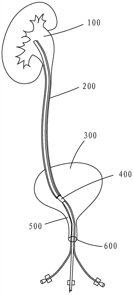
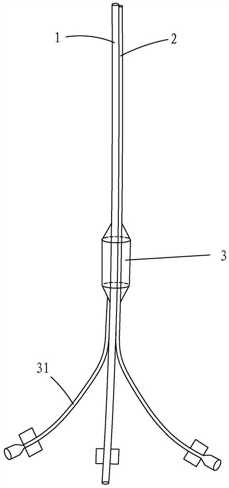
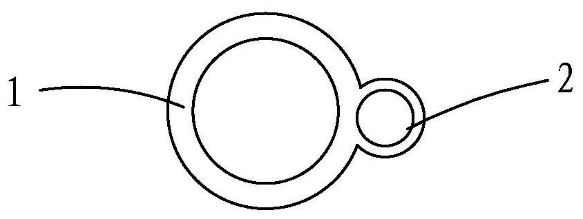
[0032] Such as Figure 1 to Figure 6 As shown, a renal pelvis pressure monitoring balloon perfusion catheter includes: a main catheter 1, a pressure measuring tube 2, and a balloon 3, the side walls of the main catheter 1 and the pressure measuring tube 2 are connected in parallel, and are disposed at the end outside the body open; the end of the pressure measuring tube 2 is connected with a transducer and a pressure monitor outside the body. The inner diameter of the main conduit 1 is larger than the inner diameter of the pressure measuring tube 2 .

[0033] The balloon 3 is wrapped around the periphery of the main catheter 1 and the piezometric tube 2 connected in parallel, and is located 30 cm above the end of the parallel section of the main catheter 1 and the piezo tube 2 ( Image 6 Shown as L in the middle); the bottom of the balloon 3 is connected to a balloon water injection tube 31, and the end of the balloon water injection tube 31 is connected to a pressure monitor...


PUM


Abstract

A renal pelvis pressure monitoring balloon perfusion catheter comprises a main catheter and a balloon. The head end of the main catheter enters from the urethral orifice of a human body and retrogradeto the renal pelvis through the urethra, the bladder, the ureter orifice and the ureter, and the tail end of the main catheter extends out of the body; the balloon wraps the periphery of the main catheter and is located in the ureter and above the ureter wall interval section when the balloon is placed in the body. The bottom of the balloon is connected with a balloon water injection pipe, and the tail end of the balloon water injection pipe is connected with a pressure monitoring injector in vitro. The catheter further comprises a pressure measuring pipe; the side walls of the piezometric tube and the main catheter are connected in parallel and are separated at the tail end in vitro; the tail end of the piezometric tube is externally connected with a transducer and a pressure monitor; the balloon wraps the periphery of the main catheter and the piezometric tube which are connected in parallel. The invention provides a renal pelvis pressure monitoring balloon perfusion catheter capable of achieving controllable and safe perfusion.

Description

【Technical field】 [0001] The invention belongs to the technical field of medical devices, and specifically refers to a renal pelvis pressure monitoring balloon perfusion catheter. 【Background technique】 [0002] In clinical practice, pelvic ureteral infusion chemotherapy is needed for patients with low-risk renal pelvis and ureteral carcinoma. Infusion chemotherapy generally requires chemotherapeutic drugs to remain in the renal collecting system (including the renal calyces, major renal calyces, and renal pelvis) and the ureter for 30 minutes. Anatomically, when the ureter passes through the bladder wall, it forms a "ureterovesical interwall segment", which is the last segment of the ureter and the smallest diameter of the ureter (the normal diameter is 2mm, and it can be expanded to 6-8mm when taking drugs to expel stones Even larger), this section can usually play the role of ureter against vesicourinary reflux, and the circumference of the ureter above this section is r...

Claims


Application Information

Patent Timeline
Patent Type & Authority Applications(China)
IPC IPC(8): A61M25/10A61M25/00A61M31/00A61B5/03A61B5/20A61B5/00
CPCA61B5/036A61B5/201A61B5/4836A61B5/4839A61B5/6853A61M25/0026A61M25/10A61M31/00A61M2025/0002A61M2025/0003A61M2025/0037A61M2205/3331A61M2205/3344A61M2210/1082A61M2210/1089A61M2230/00A61M25/10184A61M2210/005
Inventor 吴进锋叶烈夫魏永宝
Owner FUJIAN PROVINCIAL HOSPITAL
Why Eureka
Try Eureka