Animal heart simulated uterus containing pressing device and preparation method of animal heart simulated uterus
A technology of presser and uterus, which is applied in the field of medical models, can solve the problems of poor teaching effect of uterus models and the inability to simulate the reality of clinical surgical operations, so as to improve the sense of real experience, improve clinical experience, and facilitate surgical operations.
- Summary
- Abstract
- Description
- Claims
- Application Information
AI Technical Summary
Problems solved by technology
Method used
Image
Examples
preparation example Construction
[0039] The second aspect of the present invention provides a method for preparing the above-mentioned animal heart simulated uterus containing the presser 1, the preparation method at least includes the following steps:
[0040] S1. Select fresh animal hearts as raw materials. Animal hearts that are similar in size to the human uterus can be used to prepare a simulated uterus, preferably any one of pig heart, beef heart, and sheep heart;
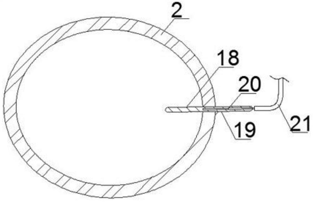
[0041] S2. Pretreating the animal heart selected in step S1 to obtain a uterine primary model 2;
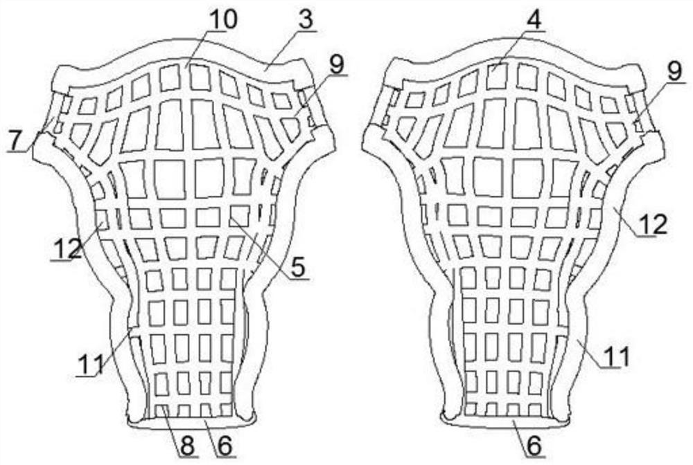
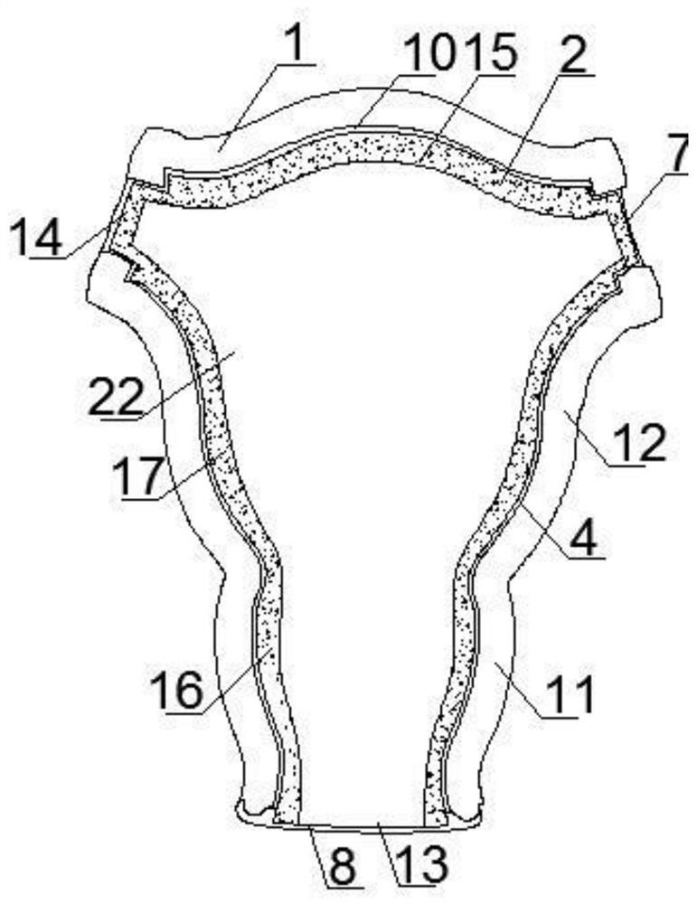
[0042] S3, put the uterine primary mold 2 obtained in step S2 into the mold cavity 5 of the lower mold 4 of the presser 1, and adjust the position of the uterine primary mold 2 in the mold cavity 5, so that the outer wall of the uterine primary mold 2 All parts are in close contact with the inner wall of the mold cavity 5;
[0043] S4. Cut off the animal heart muscle of the uterine primary mold 2 located outside the presser 1 in step S3, and k...
Embodiment 1
[0054] S1, select fresh pig heart as raw material;
[0055] S2. The pig heart selected in the step S1 is preprocessed to obtain the initial uterus model 2, as follows:
[0056] S21, connection: Use surgical scissors to cut off the chords of the pig heart and the intervals inside the pig heart, so that the cavities of the pig heart are connected;
[0057] S22. Thinning: use surgical scissors to cut away the myocardial tissue from the inner wall of the pig heart to thin the thickness of the myocardial tissue of the pig heart, and retain the outer surface of the pig heart and the myocardial tissue 1.0 cm below the outer surface of the pig heart;
[0058] S23. Overturning: turning the inner side wall of the thinned pig heart outward from the opening at the top of the pig heart, so that the outer wall of the pig heart is turned over to become the inner surface, thereby making the uterine primary mold 2;
[0059] S3. Put the primary uterine mold 2 obtained in step S2 into the mold ...
Embodiment 2
[0063] S1, select fresh pig heart as raw material;
[0064] S2. The pig heart selected in the step S1 is preprocessed to obtain the initial uterus model 2, as follows:
[0065] S21, connection: Use surgical scissors to cut off the chords of the pig heart and the intervals inside the pig heart, so that the cavities of the pig heart are connected;
[0066] S22. Thinning: Use surgical scissors to cut away the myocardial tissue from the inner wall of the pig heart outward to thin the thickness of the myocardial tissue of the pig heart, and retain the outer surface of the pig heart and the myocardial tissue 1.5 cm below the outer surface of the pig heart;
[0067] S23. Overturning: turning the inner side wall of the thinned pig heart outward from the opening at the top of the pig heart, so that the outer wall of the pig heart is turned over to become the inner surface, thereby making the uterine primary mold 2;
[0068] S3. Put the primary uterine mold 2 obtained in step S2 into t...
PUM
| Property | Measurement | Unit |
|---|---|---|
| The inside diameter of | aaaaa | aaaaa |
| The inside diameter of | aaaaa | aaaaa |
| The inside diameter of | aaaaa | aaaaa |
Abstract
Description
Claims
Application Information
 - R&D Engineer
- R&D Manager
- IP Professional
- Industry Leading Data Capabilities
- Powerful AI technology
- Patent DNA Extraction
Browse by: Latest US Patents, China's latest patents, Technical Efficacy Thesaurus, Application Domain, Technology Topic, Popular Technical Reports.
© 2024 PatSnap. All rights reserved.Legal|Privacy policy|Modern Slavery Act Transparency Statement|Sitemap|About US| Contact US: help@patsnap.com


