


Quick Research
Generate reliable direction feasibility study reports for your R&D in just a few steps.

Technical Q&A
Discover and master advanced knowledge NOW. Basics, ideas, possibilities, all at once.

Find Solutions
As an expert in R&D theories, this can generate solutions to your technical problems instantly.

Evaluate Feasibility
Analyze your overall solution with one click, know your potential R&D risks in advance.

Monitor Landscape
Get weekly tech updates, stay abreast of the latest tech innovations and key insights.
Cardiomyocyte isolation reagent and isolation method
A technology of cardiomyocytes and separation methods, applied in the direction of cell dissociation methods, biochemical equipment and methods, animal cells, etc., can solve the problems of human cardiomyopathy research and experiment limitations, can not fully simulate human physiological and pathological conditions, and achieve Improvement of cell viability and quality, shortening of operation time
- Summary
- Abstract
- Description
- Claims
- Application Information
AI Technical Summary
Problems solved by technology
Method used
Image


Examples
Embodiment 1
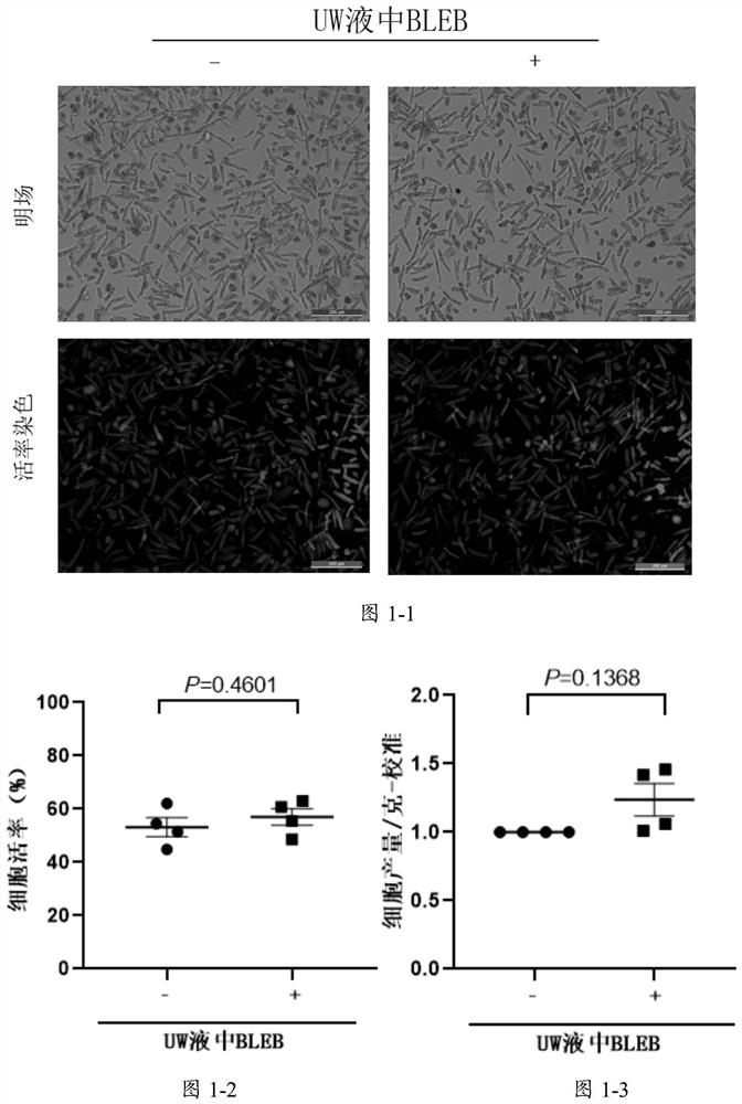
[0101] Example 1: Effect of using BLEB in the preprocessing step
[0102] In the traditional myocardial cell separation process, after the myocardial tissue is removed, the tissue is placed in organ preservation solution (UW solution) for slicing and shredding. The operation time in UW solution is about 1.5 hours. In this example, the effect of adding BLEB to the UW solution used in the traditional method on the separation effect in the pretreatment step was studied. Specifically, myocardial tissue was collected from the left atrial appendage of 4 male patients (age 75±7 years old) undergoing heart disease surgery, and the pretreatment solution was obtained by adding 10 μM BLEB to the UW solution, and the removed tissue was divided into two equally, respectively Proceed to the slicing and shredding steps. After shredding, decalcification, digestion and recalcification were performed according to the literature method (Guo et al., Amodified method for isolation of human cardio...
Embodiment 2
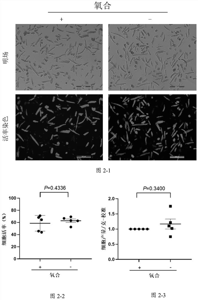
[0103] Example 2: Effect of presence or absence of an oxygenation step
[0104] First, prepare calcium-free liquid according to the following formula:
[0105] Element concentration Element concentration Sodium chloride 126mM taurine 2mM potassium chloride 4.4mM Creatine 5mM Magnesium chloride hexahydrate 5mM sodium pyruvate 5mM Sodium dihydrogen phosphate 5mM penicillin 200U / ml 4-Hydroxyethylpiperazineethanesulfonic acid 5mM streptomycin 200μg / ml glucose 22mM BLEB 10μM Deionized water
[0106] The above-mentioned calcium-free solution was divided into two groups for experimentation, and the calcium-free solution of one group was continuously filled with oxygen for 20 minutes before use (the oxygenated group), and the calcium-free solution of the other group was not filled with oxygen (the non-oxygenated solution). group), directly used for the isolation of cardiomyocytes. Myocard...
Embodiment 3
[0108] Embodiment 3: the comparison of cleaning decalcification method and gradient decalcification method
[0109]Myocardial tissues were collected from the left atrial appendages of 4 heart disease patients (aged 50-70 years old), and were equally divided into two groups after the pretreatment step of Example 1. The first group carries out the gradient decalcification operation known in the prior art, uses the calcium-free liquid used in embodiment 2 to divide and carry out gradient decalcification 3 times, and the decalcification time is 2, 3, 4 minutes respectively, during this period constantly with sequential Shake the Erlenmeyer flask clockwise to make the tissue fully contact with the calcium-free solution, so that the calcium ions in the cells in the tissue can continuously flow out. After each timing, filter out the calcium-free solution in the bottle, and replace it with a new calcium-free solution for the next decalcification. The second group did not perform the ...
PUM
 Abstract
Description
Claims
Application Information
 - R&D Engineer
- R&D Manager
- IP Professional
- Industry Leading Data Capabilities
- Powerful AI technology
- Patent DNA Extraction
Browse by: Latest US Patents, China's latest patents, Technical Efficacy Thesaurus, Application Domain, Technology Topic, Popular Technical Reports.
© 2024 PatSnap. All rights reserved.Legal|Privacy policy|Modern Slavery Act Transparency Statement|Sitemap|About US| Contact US: help@patsnap.com