
A medical ultrasonic scanning device with adjustable pressure based on a mechanical arm

A scanning device and adjustable pressure technology, applied in the field of medical robots, can solve the problems of patients' skin tissue damage, easy fatigue, and poor scanning images obtained by the probe

Active Publication Date: 2021-08-20
ZHEJIANG UNIV
View PDF16 Cites 0 Cited by
  • Summary
  • Abstract
  • Description
  • Claims
  • Application Information

AI Technical Summary

Problems solved by technology

The existing robot-assisted ultrasonic scanning equipment that has been developed adopts mechanical scanning, which overcomes the shortcomings of manual scanning, such as fatigue, hand shaking, and unstable pressure, which affect the quality of ultrasonic imaging. However, the ultrasonic probe is usually fixed in a simple way. Rigid fixation mainly has the following problems: 1. During the mechanical scanning process, the patient’s skin tissue damage or even danger may be caused due to unreceived feedback or feedback delay; 2. Due to the complexity of the patient’s body (back curve) shape) and tissue deformation, it is difficult to collect effective information to control and adjust the probe; 3. If the probe is not adjusted in time, a better scan image may not be obtained

Method used


Image

  • A medical ultrasonic scanning device with adjustable pressure based on a mechanical arm
  • A medical ultrasonic scanning device with adjustable pressure based on a mechanical arm
  • A medical ultrasonic scanning device with adjustable pressure based on a mechanical arm

Examples


Embodiment 1

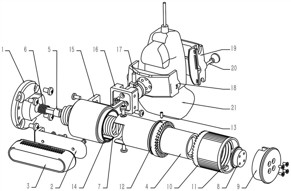
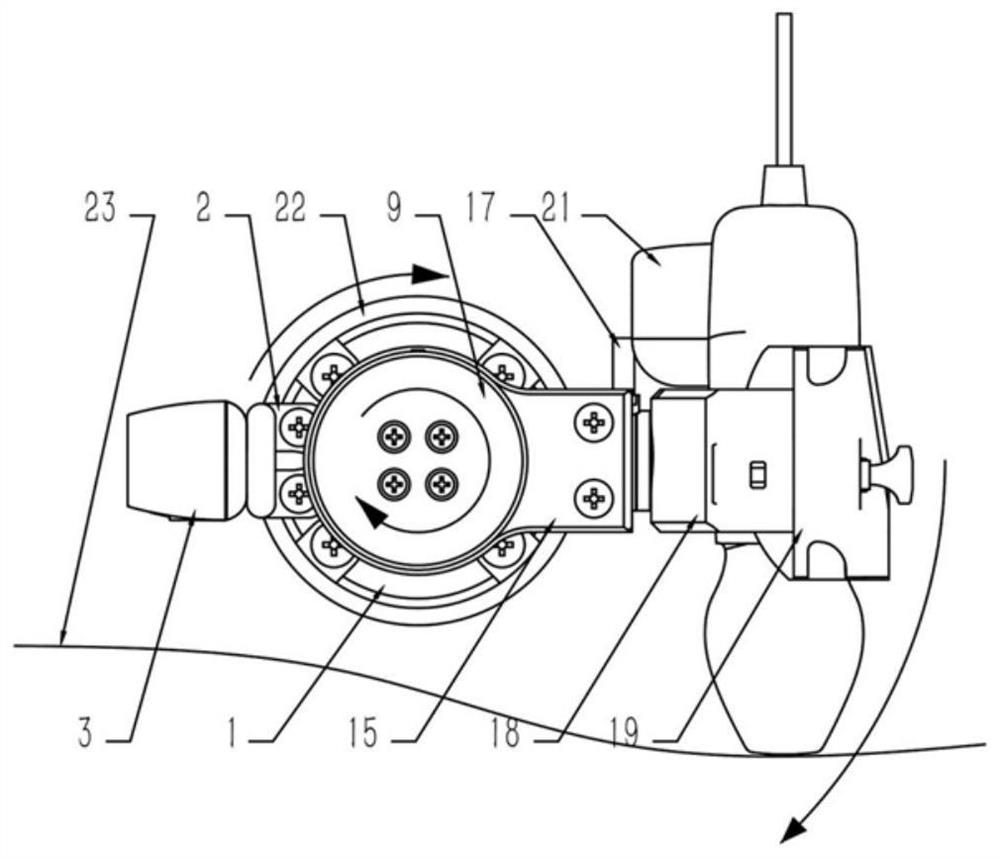
[0051] The entire ultrasonic scanning device is fixed on the end flange 22 of the UR5 (a collaborative robot arm produced by Universal Robots) through four mounting holes distributed in a circular array on the base 1, for scanning the spine on the back of the human body. imaging.

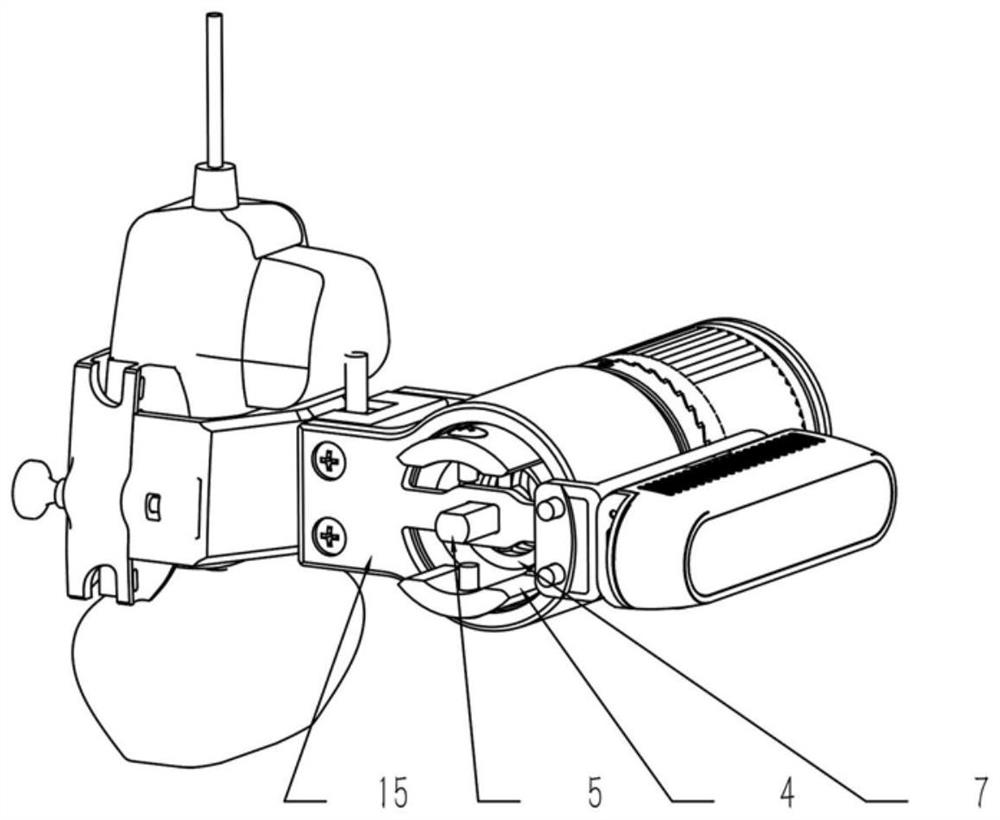
[0052] The hospital selects a suitable probe fixing block 19 according to the straight probe or curved surface probe used, and places the probe 21 on the probe base 18, and the rear side is close to the curved surface of the probe base. The probe is wrapped with waterproof tape to eliminate fit gaps and improve grip. Then align the probe fixing block 19 with the through hole of the probe base 18, and insert the hand-twist bolt to rotate and fix it. Note that the notch at the end of the through hole on both sides of the probe base 18 can be inserted into the nut. It is not necessary to fully rotate the bolt when disassembling the probe. It can be unscrewed with a handle attached to the nut of the bo...

Embodiment 2

[0057] The present invention sets a triaxial force sensor to collect the pressure loaded on the human body by the ultrasonic probe 21 . Developers of medical ultrasound scanning systems can also develop force control programs to control the rotation of the flange at the end of the mechanical arm according to the pressure to achieve constant pressure control. The characteristic curve of the torsion spring makes the rigidity curve smooth during the scanning process, providing support for constant force control.

Embodiment 3

[0059] The present invention is equipped with a Realsense depth camera 3, and the developer of the medical ultrasound scanning system can also control the automatic operation of the mechanical arm according to the point cloud data obtained by the camera, and move to scan above the human body.


PUM


Abstract

The invention discloses a medical ultrasonic scanning device with adjustable pressure based on a mechanical arm. The device includes three parts: a camera bracket, a torque adjustment mechanism and a probe rotating arm. The invention is installed on a cooperative mechanical arm with no less than six degrees of freedom. On the end flange, the torsion spring is used to drive the probe to rotate to realize that the ultrasonic probe is loaded on the human body to obtain ultrasonic images and move for scanning. The torsion spring of the present invention isolates the rigid loading of the human body by the scanning operation of the mechanical arm, on the one hand avoids the impact damage to the human body caused by rigid contact, and on the other hand adjusts the rigidity curve of the loading process to provide support for voltage stabilization control. Finally, the device is equipped with a three-axis force sensor and RGB-D camera, which provides a solution for force control and visual servo ultrasound scanning to ensure the safety, reliability and high-quality imaging of robot-assisted medical ultrasound scanning.

Description

technical field [0001] The invention relates to the field of medical robots, and is subdivided into the field of medical ultrasonic scanning, in particular to robot-assisted ultrasonic scanning of the human spine, and in particular to a medical ultrasonic scanning device with pressure regulating capability. Background technique [0002] The traditional medical ultrasound scan method is a hand-held manual ultrasound scan. The doctor holds an ultrasound probe to scan the corresponding body parts of the patient to obtain the three-dimensional structural characteristics of the patient's examination site. The existing robot-assisted ultrasonic scanning equipment that has been developed adopts mechanical scanning, which overcomes the shortcomings of manual scanning, such as fatigue, hand shaking, and unstable pressure, which affect the quality of ultrasonic imaging. However, the ultrasonic probe is usually fixed in a simple way. Rigid fixation mainly has the following problems: 1....

Claims


Application Information

Patent Timeline
Patent Type & Authority Patents(China)
IPC IPC(8): A61B8/08A61B8/00B25J17/00B25J18/00
CPCA61B8/085A61B8/0875A61B8/4209A61B8/4444A61B2562/0247B25J17/00B25J18/00
Inventor 杨克己王云江朱永坚
Owner ZHEJIANG UNIV
Learn More