
Magnetic resonance imaging method and device and storage medium

A magnetic resonance imaging and pulse preparation technology, which is applied in the field of medical imaging, can solve the problems of impact, scanning time and resolution limitations, and the inability to obtain magnetic resonance images, etc., and achieve the effect of high contrast and spatial resolution and high navigation efficiency

Active Publication Date: 2020-09-08
TSINGHUA UNIV
View PDF5 Cites 3 Cited by
  • Summary
  • Abstract
  • Description
  • Claims
  • Application Information

AI Technical Summary

Problems solved by technology

Since the heart will be affected by its own movement and respiratory movement, breath-holding is generally used in existing imaging sequences, so that the scanning time and resolution will be limited, and the imaging scanning time based on the existing breathing navigation method will also be limited. Impact of Breath Navigation Efficiency
Therefore, ideal MRI images cannot be obtained

Method used


Image

  • Magnetic resonance imaging method and device and storage medium
  • Magnetic resonance imaging method and device and storage medium
  • Magnetic resonance imaging method and device and storage medium

Examples


Embodiment Construction

[0064] In order to make the objects, technical solutions and advantages of the present invention more apparent, exemplary embodiments according to the present invention will be described in detail below with reference to the accompanying drawings. Apparently, the described embodiments are only some embodiments of the present invention, rather than all embodiments of the present invention, and it should be understood that the present invention is not limited by the exemplary embodiments described here. Based on the embodiments of the present invention described in the present invention, all other embodiments obtained by those skilled in the art without creative effort shall fall within the protection scope of the present invention.

[0065] In order to obtain images with higher contrast and spatial resolution, an embodiment of the present invention provides a magnetic resonance imaging method. The magnetic resonance imaging method can be applied to various target tissues such a...


PUM


Abstract

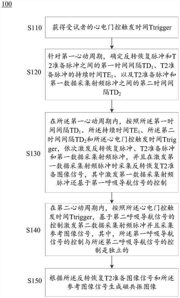
The invention provides a magnetic resonance imaging method and device and a storage medium. The method comprises the following steps: obtaining electrocardiograph gating trigger time Ttrigger; for thefirst cardiac cycle, determining a time interval TD1 between inversion recovery pulse and T2 preparation pulse, a duration TE1 of the T2 preparation pulse, and a time interval TD2 between the T2 preparation pulse and a first data acquisition radio frequency pulse; in the first cardiac cycle, exciting the pulses according to the TD1, the TE1, the TD2 and the Ttrigger, and collecting inversion recovery T2 preparation image signal when the first data collection radio frequency pulse is excited, wherein the excitation of the first data collection radio frequency pulse is further based on controlof a respiratory navigation signal; in a second cardiac cycle, exciting a second data acquisition radio frequency pulse based on the control of the respiratory navigation signal according to the Ttrigger, and acquiring a reference image signal, wherein the control of the respiratory navigation signal in the two cycles is independent; and generating a magnetic resonance image according to the two image signals. According to the scheme, the contrast and the spatial resolution of the image can be remarkably improved.

Description

technical field [0001] The present invention relates to the field of medical imaging, and more specifically relates to a magnetic resonance imaging method, device and storage medium. Background technique [0002] Magnetic resonance imaging technology uses the phenomenon of nuclear magnetic resonance to image the human body. It is already a common medical imaging examination method and can be applied to various medical application scenarios. For example, atrial fibrillation is the most common arrhythmia in clinical practice, and radiofrequency ablation of the left atrium and pulmonary vein isolation are the main treatments for controlling atrial fibrillation. The degree of atrial fibrosis and scar tissue after radiofrequency ablation are related to the recurrence of atrial fibrillation. Therefore, atrial fibrosis and the anatomical structure of surrounding tissues can help decision-making before ablation, and the scar damage produced by ablation can also reflect the prognosis...

Claims


Application Information

Patent Timeline
IPC IPC(8): A61B5/055
CPCA61B5/055
Inventor 丁海艳司东岳
Owner TSINGHUA UNIV
Learn More