Method for detecting standard uptake value of thoracic lymph node based on CT imaging omics

A standard uptake value and CT image technology, applied in the field of medical image processing and analysis, can solve the problems of increasing medical costs and patient burdens

Pending Publication Date: 2020-08-28
JILIN UNIV FIRST HOSPITAL
View PDF4 Cites 0 Cited by
  • Summary
  • Abstract
  • Description
  • Claims
  • Application Information

AI Technical Summary

Problems solved by technology

However, PET / CT, as a high-end inspection method, greatly increases the cost of medical care and the burden on patients

Method used


Image

  • Method for detecting standard uptake value of thoracic lymph node based on CT imaging omics
  • Method for detecting standard uptake value of thoracic lymph node based on CT imaging omics
  • Method for detecting standard uptake value of thoracic lymph node based on CT imaging omics

Examples


Embodiment Construction

[0031] The present invention will be further described in detail below in conjunction with the accompanying drawings, so that those skilled in the art can implement it with reference to the description.

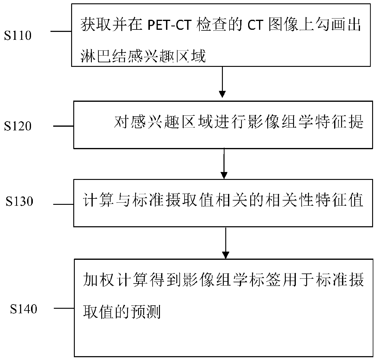
[0032] The radiomics-based detection method of the chest lymph node standard uptake value provided by the present invention includes:

[0033] Step S110, obtain the patient's PET-CT examination image, and outline the region of interest on the CT image of the PET-CT examination; wherein, the region of interest is manually drawn by the doctor, which is the mediastinal and hilar lymph nodes in the PET-CT examination image your region.

[0034] Step S120, perform feature extraction on the region of interest, and obtain the corresponding edge features and region features in the region of interest; wherein, the edge features include: edge feature parameters and discrete cosine transform parameters composed of discrete cosine transform sub-coefficients independent moment invariant p...


PUM


Abstract

The invention provides a method for detecting A standard uptake value of A thoracic lymph node based on CT imaging omics, which comprises the following steps: step 1, acquiring a PET-CT examination image of a patient, and drawing a lymph node region of interest on a CT image in the PET-CT examination image; step 2, carrying out image omics feature extraction on the region of interest, wherein theimage omics feature extraction comprises edge features and region features; step 3, preprocessing the edge features and the region features, and step 4, carrying out weighting processing on the feature values with correlation, and carrying out linear summation to obtain an imaging omics label for predicting a standard uptake value so as to be used for predicting benign and malignant lymph nodes. According to the invention, a lymph node region of interest is manually drawn; and feature extraction is performed on the region of interest, features highly related to the standard uptake value are screened through correlation analysis, and linear weighting calculation is performed to obtain an imaging omics label for predicting the standard uptake value.

Description

technical field [0001] The invention relates to the field of processing and analysis of medical images, in particular to a method for detecting standard uptake values ​​of chest lymph nodes based on radiomics. Background technique [0002] Lung cancer is a malignant tumor with increasing incidence rate, and the rising trend is obvious in developing countries. In China, the morbidity and mortality of lung cancer rank first among malignant tumors. Statistics show that in 2013, about 530,700 lung cancer patients died in my country, and the number of new lung cancer cases reached 732,800. The number of new cases and deaths The number accounts for 19.90% and 26.20% of all malignant tumors in the country, ranking first among malignant tumors. At present, 70% to 80% of lung cancer patients in clinical practice are in the middle and advanced stages when they are diagnosed, and the incidence of lymph node metastasis is high. The location and number of lymph node metastasis play a dec...

Claims


Application Information

Patent Timeline
IPC IPC(8): G06T7/00G06T7/11G06T7/13G06F17/14
CPCG06T7/0012G06T7/13G06T7/11G06F17/147G06T2207/10081G06T2207/10104G06T2207/20104G06T2207/30061G06T2207/30096
Inventor 张惠茅张磊付宇杨琪谢云明
Owner JILIN UNIV FIRST HOSPITAL
Why Eureka
Try Eureka