
Medical image visualization method and device, equipment and computer storage medium

A medical image and image technology, applied in medical image, computing, image enhancement, etc., can solve the problems of internal detail information loss, noise not improved, medical image tomography effect, etc., to reduce the loss of internal detail information and weaken the fault Effects, Effects that Improve Display Effects

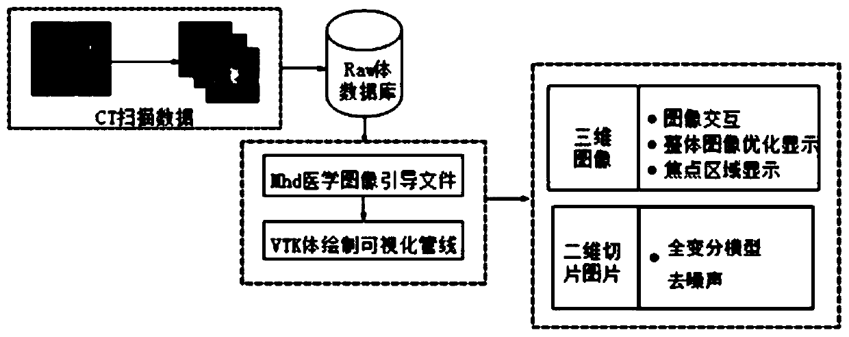
Pending Publication Date: 2020-08-14
中移雄安信息通信科技有限公司 +1
View PDF3 Cites 0 Cited by
  • Summary
  • Abstract
  • Description
  • Claims
  • Application Information

AI Technical Summary

Problems solved by technology

However, this method is based on the traditional gradient (segmented) transfer function to realize the visualization of medical images, so that the displayed medical image tomography effect is obvious, the internal details are seriously lost, and the noise problem has not been improved.

Method used


Image

  • Medical image visualization method and device, equipment and computer storage medium
  • Medical image visualization method and device, equipment and computer storage medium
  • Medical image visualization method and device, equipment and computer storage medium

Examples


Embodiment Construction

[0046] The characteristics and exemplary embodiments of various aspects of the present invention will be described in detail below. In order to make the purpose, technical solutions and advantages of the present invention clearer, the present invention will be further described in detail below in conjunction with the accompanying drawings and specific embodiments. It should be understood that the specific embodiments described here are only configured to explain the present invention, not to limit the present invention. It will be apparent to one skilled in the art that the present invention may be practiced without some of these specific details. The following description of the embodiments is only to provide a better understanding of the present invention by showing examples of the present invention.

[0047] It should be noted that in this article, relational terms such as first and second are only used to distinguish one entity or operation from another entity or operation...


PUM


Abstract

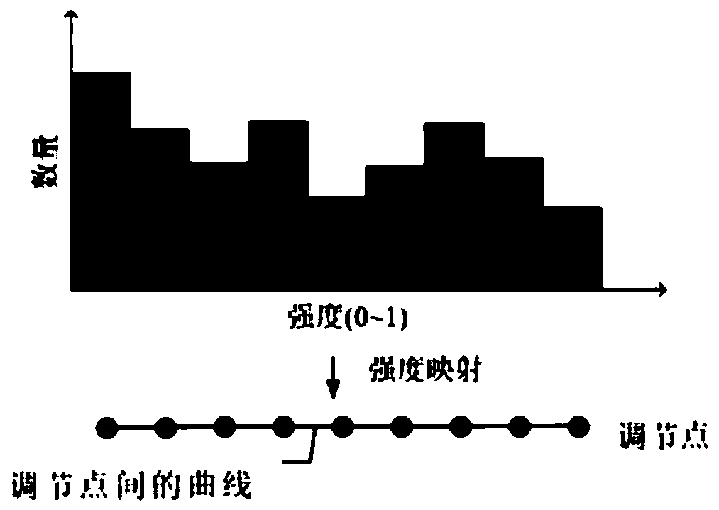
The embodiment of the invention discloses a medical image visualization method and device, equipment and a computer storage medium. The visualization method of the medical image comprises the following steps: obtaining a first three-dimensional medical image based on acquired medical image volume data; determining a first volume data intensity value of each piece of medical image volume data; based on a preset semi-automatic transfer function and the first volume data intensity value of each piece of medical image volume data, determining a first opacity value corresponding to each piece of medical image volume data, wherein the semi-automatic transfer function represents a mapping relationship between the volume data intensity value and the opacity value; and based on the first opacity value corresponding to each piece of medical image volume data, adjusting the first three-dimensional medical image to obtain a second three-dimensional medical image. According to the embodiment of theinvention, the display effect of the medical image can be improved.

Description

technical field [0001] The invention belongs to the technical field of image visualization, and in particular relates to a medical image visualization method, device, electronic equipment and computer storage medium. Background technique [0002] Visualization is a technology that converts data into images and presents them on the screen through computer graphics and graphics processing technology, and can further perform interactive operations, thus giving birth to medical image visualization technology. [0003] At present, the common three-dimensional visualization method of medical images: medical image generation is through the integration of QT interface and VTK (visualization toolkit), reading data source, image materialization, three-dimensional reconstruction, rendering and display and other steps. However, this method is based on the traditional gradient (segmented) transfer function to realize the visualization of medical images. The medical image displayed in thi...

Claims


Application Information

Patent Timeline
Patent Type & Authority Applications(China)
IPC IPC(8): G06T17/00G06T5/00G16H30/40
CPCG06T17/00G16H30/40G06T2207/10081G06T5/70
Inventor 魏敏
Owner 中移雄安信息通信科技有限公司
Learn More