
Method of detecting protein complex in biological sample

A detection method and complex technology, applied in the fields of biochemical analysis and protein analysis, can solve problems such as lack of antibodies, and achieve the effect of enriching sources and reducing steric hindrance effects.

Pending Publication Date: 2020-05-22
BGI CLINICAL LAB (SHENZHEN) CO LTD
View PDF4 Cites 1 Cited by
  • Summary
  • Abstract
  • Description
  • Claims
  • Application Information

AI Technical Summary

Problems solved by technology

[0007] The purpose of the present invention is to solve the problem of steric hindrance after using chemical cross-linking agents to stabilize protein complexes and the lack of antibodies for co-immunoprecipitation when cross-linking mass spectrometry and co-immunoprecipitation techniques are combined in the prior art. It is beneficial to carry out the study of protein complex and protein interaction

Method used


Image

  • Method of detecting protein complex in biological sample
  • Method of detecting protein complex in biological sample
  • Method of detecting protein complex in biological sample

Examples


Embodiment 1

[0052] In this embodiment, Hela cells are taken as an example, heat shock protein 90 is used as the target protein, and the heat shock protein complex is detected in situ in the cell, and the specific operation is as follows.

[0053] (1) Covalent crosslinking

[0054] Will 1×10 7 Resuspend Hela cells in 450 μl phosphate buffer, add 50 μl 50 mM DSSO stock solution to make the final concentration 5 mM, place at 4 ° C for 1 h, add 1 M NH 4 HCO 3 The final concentration was 20mM to terminate the reaction, and centrifuged at 500×g for 5min to obtain cross-linked cells.

[0055] (2) Denaturation treatment

[0056] Divide the cross-linked cells into two parts, directly add 8M urea lysate 3 times the volume of the cells to one part of the cross-linked cells, sonicate on ice for 5 minutes (on 2s and stop for 3s), and extract denatured protein to obtain denatured protein solution (hereinafter Referred to as the treatment group according to the method of the present invention), anot...

Embodiment 2

[0066] In this embodiment, A549 cells are taken as an example, glyceraldehyde-3-phosphate dehydrogenase (GAPDH) is used as the target protein, and the GAPDH protein complex is detected in the cell lysate, and the specific operation is as follows.

[0067] (1) Covalent crosslinking

[0068] Will 1×10 7 A549 cells were resuspended in 450 μl phosphate buffer, added non-denaturing cell lysate (25mM HEPES, 150mM NaCl, 1% Triton X-100, pH 7.4) to extract the whole protein solution of the cells, and quantitatively obtained 4.4mg / ml protein solution 0.5 ml. Then add 50mM DSBU stock solution to make the final concentration 5mM, place it at 4°C for 1h, add 1M NH 4 HCO 3 The final concentration was 20mM to stop the reaction, and centrifuged at 500×g for 5min to obtain the cross-linked protein solution.

[0069] (2) Denaturation treatment

[0070] The cross-linked protein solution was divided into two parts and diluted to 2.2 mg / ml with non-denaturing cell lysate (25 mM HEPES, 150 mM...


PUM


Abstract

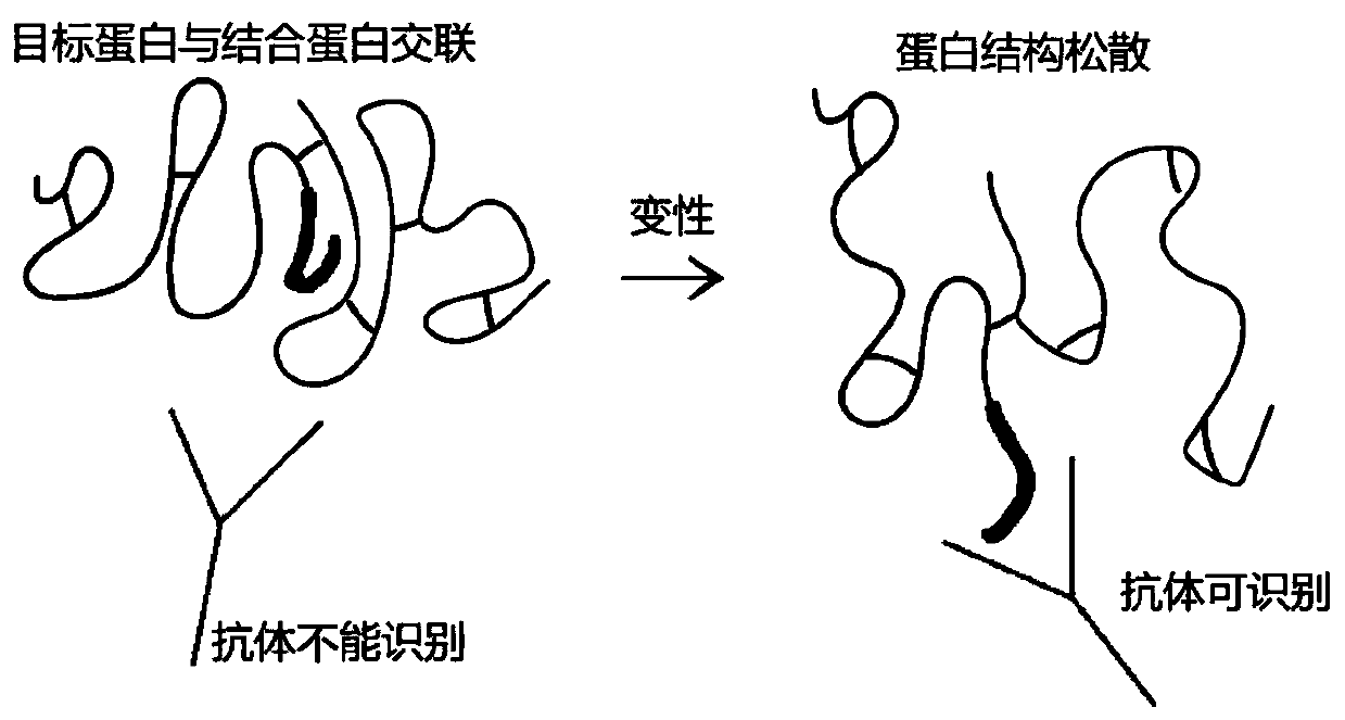
The invention provides a method for detecting a protein complex in a biological sample. The method comprises the steps of: covalent crosslinking, denaturation treatment, solution replacement, co-immunoprecipitation and mass spectrometry. According to the method, a three-dimensional structure of protein is destroyed through the denaturation treatment step to disperse a peptide chain, so that the steric hindrance effect of the peptide chain is reduced, and the protein complex can be better recognized by a specific antibody. Moreover, due to denaturation treatment, an antigenic determinant buriedin a hydrophobic center of the peptide chain is exposed to serve as a linear epitope, an antibody directed at the linear epitope can be adopted for co-immunoprecipitation, and a novel thought is provided for co-immunoprecipitation. The detection method disclosed by the invention is beneficial to research on the interaction of the protein complex and the protein.

Description

technical field [0001] The invention belongs to the technical field of biochemical analysis, in particular to the technical field of protein analysis, and specifically relates to a method for detecting protein complexes in biological samples, which can study protein interaction. Background technique [0002] There are many proteins (including enzymes) in organisms, and the interactions between various proteins and proteins play an important role in maintaining the normal physiological functions of organisms. Protein interaction refers to the process in which two or more protein molecules form a protein complex through non-covalent bonds. Protein interaction involves almost all life activities, including DNA replication and transcription, protein synthesis and secretion, signal transduction and metabolism, and so on. Moreover, many protein molecules with important functions often function in the form of dimers, trimers or oligomers, and the interaction between these proteins...

Claims


Application Information

Patent Timeline
Patent Type & Authority Applications(China)
IPC IPC(8): G01N27/62
CPCG01N33/6848
Inventor 张可人任艳张宇星林志龙陈秋实李思奇潘火珍王欣然张元良
Owner BGI CLINICAL LAB (SHENZHEN) CO LTD
Learn More