Three-dimensional lung automatic segmentation and left and right lung separation method and system based on CT image

A technology for automatic segmentation of CT images, applied in image analysis, image enhancement, image data processing, etc., can solve problems such as difficult to obtain a 3D model of separation of the left and right lungs, difficult to process blood vessels, and difficult to separate

Active Publication Date: 2020-05-19
SHENZHEN YORATAL DMIT
View PDF8 Cites 4 Cited by
  • Summary
  • Abstract
  • Description
  • Claims
  • Application Information

AI Technical Summary

Problems solved by technology

[0003] The mainstream method usually uses a region growing algorithm based on grayscale information. This method requires the user to provide one or several seed points and set a grayscale threshold. Usually, this method will inevitably bring out the bronchi, and the left and right lungs will be connected to the Together, it is difficult to separate, the time cost of manual separation is high, and the blood vessels entering the lungs are not easy to handle
Therefore, it is usually difficult to obtain a good three-dimensional model of the separation of the left and right lungs.
[0004] In summary, there are many problems in the actual use of existing methods, so it is necessary to improve

Method used


Image

  • Three-dimensional lung automatic segmentation and left and right lung separation method and system based on CT image
  • Three-dimensional lung automatic segmentation and left and right lung separation method and system based on CT image
  • Three-dimensional lung automatic segmentation and left and right lung separation method and system based on CT image

Examples


Embodiment Construction

[0054] In order to make the object, technical solution and advantages of the present invention clearer, the present invention will be further described in detail below in conjunction with the accompanying drawings and embodiments. It should be understood that the specific embodiments described here are only used to explain the present invention, not to limit the present invention.

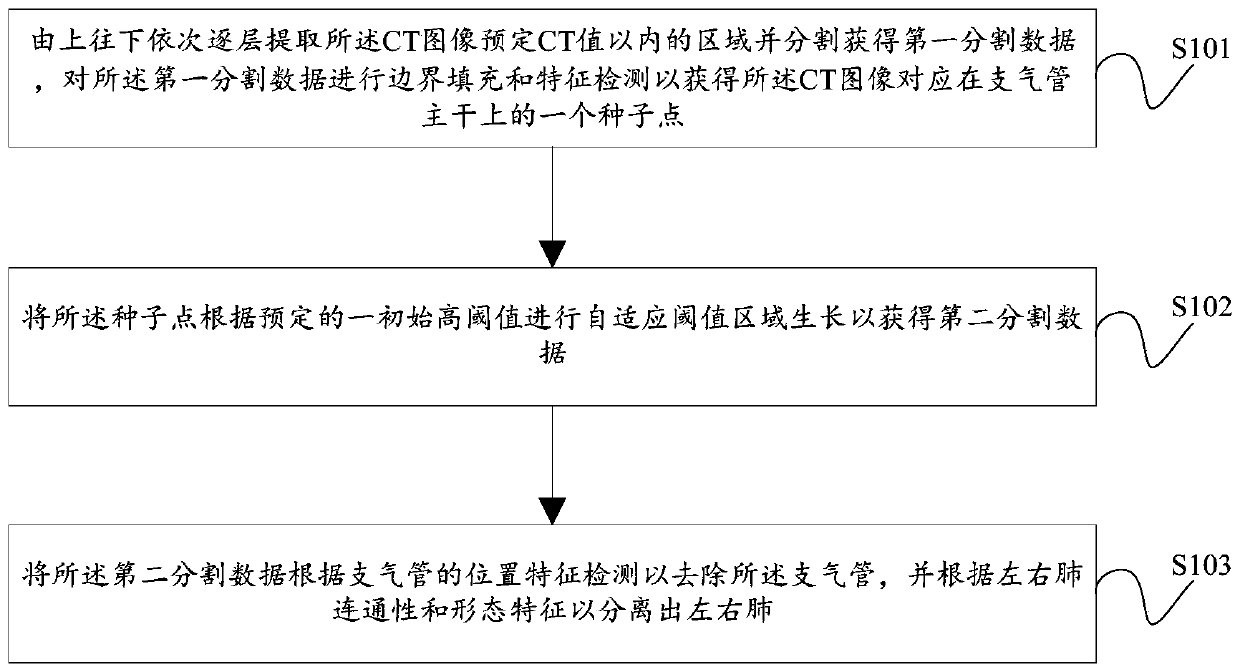
[0055] figure 1 The method for automatic three-dimensional lung segmentation and left and right lung separation based on CT images described in the embodiment of the present invention includes the following steps:

[0056] S101: Extract the region within the predetermined CT value of the CT image layer by layer from top to bottom and segment to obtain the first segmentation data, and perform boundary filling and feature detection on the first segmentation data to obtain the CT image corresponding to the bronchi A seed point on the trunk;

[0057] S102: Perform adaptive threshold region growth on ...


PUM


Abstract

The method is suitable for the technical field of computer medical image processing, and provides a three-dimensional lung automatic segmentation and left and right lung separation method based on a CT image, and the method comprises the steps: extracting regions within a preset CT value of the CT image layer by layer from the top to the bottom, and sequentially segmenting the interior and exterior of each layer of region, so as to obtain first segmentation data; sequentially carrying out boundary filling and feature detection on the first segmentation data to obtain a seed point, corresponding to a bronchial trunk, of the CT image; performing adaptive threshold region growth on the seed point according to a predetermined initial high threshold to obtain second segmentation data; and detecting the second segmentation data according to the position characteristics of the bronchus to remove the bronchus, and separating the left lung and the right lung according to the connectivity and morphological characteristics of the left lung and the right lung. The invention further provides a three-dimensional lung automatic segmentation and left and right lung separation system based on the CT image. Therefore, the left lung and the right lung of the CT image can be automatically segmented and separated, the precision is high, and the accuracy is high.

Description

technical field [0001] The invention relates to the technical field of computer medical image processing, in particular to a method and system for automatic three-dimensional lung segmentation and separation of left and right lungs based on CT images. Background technique [0002] Lung cancer has become the leading cause of cancer death worldwide. Surgical resection of the cancerous lung lobe is the treatment of choice for lung cancer. Compared with doctors directly interpreting two-dimensional CT (Computed Tomography, computerized tomography) images to formulate lung cancer surgery plans, using computers to analyze lung CT images and provide three-dimensional visualization of lung cavities has an absolute advantage in surgical planning. It plays an extremely important role in research and clinical application. Accurately segmenting the left and right lungs is the first step. [0003] The mainstream method usually uses a region growing algorithm based on grayscale informa...

Claims


Application Information

Patent Timeline
Patent Type & Authority Applications(China)
IPC IPC(8): G06T7/187G06T7/136G06T17/10G06T7/00
CPCG06T7/187G06T7/136G06T17/10G06T7/0012G06T2207/10081G06T2207/30061G06T2207/30096
Inventor 黄言松伍亚军
Owner SHENZHEN YORATAL DMIT
Why Eureka
Try Eureka