Fluorescent microsphere and antibody coupling method
A fluorescent microsphere and coupling technology, applied in the field of biological analysis, can solve the problems of low coupling efficiency and coupling strength of fluorescent microspheres and antibodies, and inappropriate labeling methods, so as to improve the coupling efficiency and improve the coupling efficiency. and coupling strength, the effect of avoiding agglutination
- Summary
- Abstract
- Description
- Claims
- Application Information
AI Technical Summary
Problems solved by technology
Method used
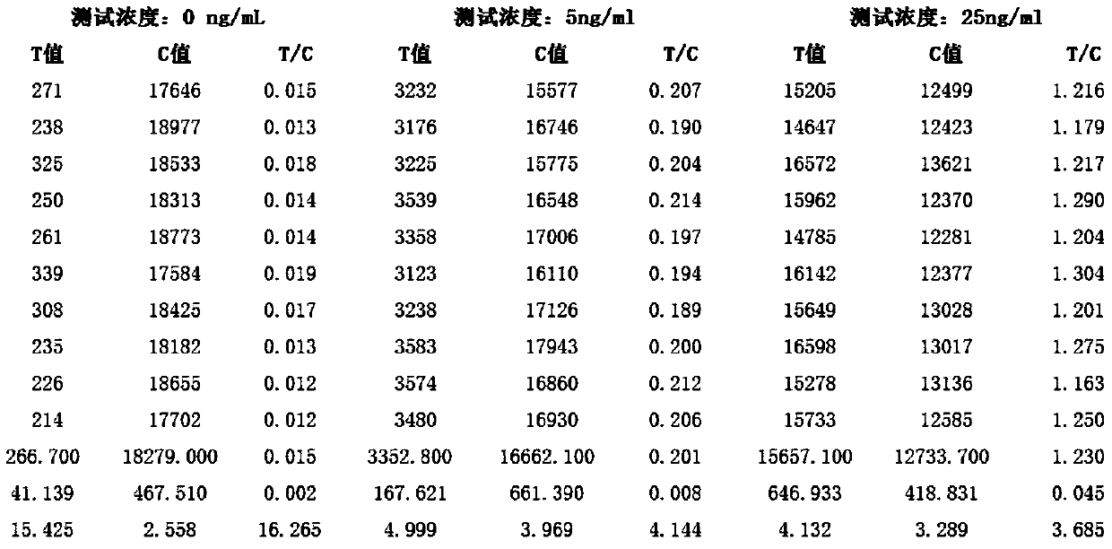
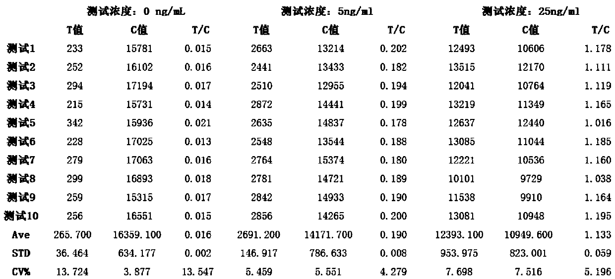
Image

Examples
Embodiment 1
[0034] This embodiment is a coupling method of fluorescent microspheres and creatine kinase isoenzyme monoclonal antibody, the coupling method comprising the following steps:
[0035] (a) After washing the fluorescent microspheres, ultrasonically disperse them for 3 minutes, dilute them with MES buffer solution with a concentration of 2% polyvinylpyrrolidone until the solid content of the fluorescent microspheres is 0.8‰, and then add them to the diluted fluorescent microsphere solution in sequence Ethanol solution containing NHS and ethanol solution containing EDC, mix well, react at room temperature for 25min, sonicate, centrifuge at 10000r / min for 10min, collect activated fluorescent microspheres, add 0.2% MES buffer solution of polyvinylpyrrolidone, ultrasonic treatment, the microsphere suspension that obtains the solid content of fluorescent microspheres is 0.08%, wherein, the addition amount of the ethanol solution containing NHS and the ethanol solution containing EDC is...
Embodiment 2
[0040] This embodiment is a coupling method of fluorescent microspheres and creatine kinase isoenzyme monoclonal antibody, the coupling method comprising the following steps:
[0041] (a) After cleaning the fluorescent microspheres, ultrasonically disperse them for 2 minutes, dilute them with MES buffer solution with a concentration of 0.05% polyvinylpyrrolidone until the solid content of the fluorescent microspheres is 1.2‰, and then add Ethanol solution containing NHS and ethanol solution containing EDC, mix well, react at room temperature for 35min, sonicate, centrifuge at 10000r / min for 10min, collect activated fluorescent microspheres, add 0.2% MES buffer solution of polyvinylpyrrolidone, ultrasonic treatment, the microsphere suspension that obtains the solid content of fluorescent microsphere is 0.12%, wherein, the addition amount of the ethanol solution containing NHS and the ethanol solution containing EDC are respectively the same as that of the fluorescent microsphere...
Embodiment 3
[0046] This embodiment is a coupling method of fluorescent microspheres and creatine kinase isoenzyme monoclonal antibody, the coupling method comprising the following steps:
[0047](a) After cleaning the fluorescent microspheres, ultrasonically disperse them for 2.5 minutes, dilute them with MES buffer solution with a concentration of 1% polyvinylpyrrolidone until the solid content of the fluorescent microspheres is 1‰, and then pour them into the diluted fluorescent microsphere solution in turn Add the ethanol solution containing NHS and the ethanol solution containing EDC, mix well, react at room temperature for 30 minutes, sonicate, centrifuge at 10000r / min for 10 minutes, collect the activated fluorescent microspheres, add 0.2 MES buffer solution of % polyvinylpyrrolidone, ultrasonic treatment, the microsphere suspension that obtains the solid content of fluorescent microspheres is 0.1%, wherein, the addition amount of the ethanol solution containing NHS and the ethanol s...
PUM
| Property | Measurement | Unit |
|---|---|---|
| particle diameter | aaaaa | aaaaa |
Abstract
Description
Claims
Application Information
 - R&D
- Intellectual Property
- Life Sciences
- Materials
- Tech Scout
- Unparalleled Data Quality
- Higher Quality Content
- 60% Fewer Hallucinations
Browse by: Latest US Patents, China's latest patents, Technical Efficacy Thesaurus, Application Domain, Technology Topic, Popular Technical Reports.
© 2025 PatSnap. All rights reserved.Legal|Privacy policy|Modern Slavery Act Transparency Statement|Sitemap|About US| Contact US: help@patsnap.com