
Fundus OCT image recognition method and device

An image recognition and image technology, applied in eye testing equipment, image enhancement, image analysis and other directions, can solve the problems of small clinical application value and poor practicability, and achieve the effect of improving the efficiency of reading images and strong practicability

Pending Publication Date: 2019-12-10
SHANGHAI EAGLEVISION MEDICAL TECH CO LTD
View PDF4 Cites 13 Cited by
  • Summary
  • Abstract
  • Description
  • Claims
  • Application Information

AI Technical Summary

Problems solved by technology

The existing technology can use the neural network to classify OCT images to obtain the identification result of a certain disease. This scheme has certain accuracy, but the clinical application value of this identification result is small and the practicability is poor.

Method used


Image

  • Fundus OCT image recognition method and device
  • Fundus OCT image recognition method and device
  • Fundus OCT image recognition method and device

Examples


Embodiment Construction

[0037] The technical solutions of the present invention will be clearly and completely described below in conjunction with the accompanying drawings. Apparently, the described embodiments are some of the embodiments of the present invention, but not all of them. Based on the embodiments of the present invention, all other embodiments obtained by persons of ordinary skill in the art without making creative efforts belong to the protection scope of the present invention.

[0038] In the description of the present invention, it should be noted that the terms "center", "upper", "lower", "left", "right", "vertical", "horizontal", "inner", "outer" etc. The indicated orientation or positional relationship is based on the orientation or positional relationship shown in the drawings, and is only for the convenience of describing the present invention and simplifying the description, rather than indicating or implying that the referred device or element must have a specific orientation, ...


PUM


Abstract

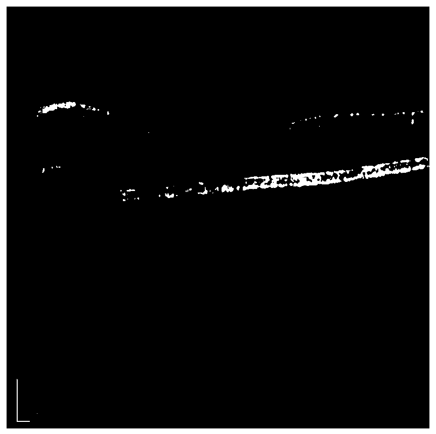
The invention provides a fundus OCT image recognition method and device, and the method comprises the steps: carrying out the recognition of a fundus OCT image through a neural network, and obtaininga recognition result related to lesion and fovea centralis; determining a region of interest according to the identification result; and determining the lesion degree according to the position and / orsize of the region of interest.

Description

technical field [0001] The invention relates to the field of ophthalmology image detection, in particular to a fundus OCT image recognition method and equipment. Background technique [0002] Machine learning technology has been widely used in the medical field, especially machine learning technology represented by deep learning has been widely concerned in the field of medical imaging. In terms of fundus image detection and differential diagnosis of eye diseases, deep learning technology has been used to detect single diseases such as glaucoma and diabetic retinopathy, and achieved good results. [0003] There are many types and large numbers of diseases that can be indicated by eye pictures, and the manifestations of the lesions are very different, ranging from large-scale fundus hemorrhage to very small hemorrhage and exudation. The diseases that can be indicated by fundus images include retinopathy (including diabetic retinopathy and other diseases), arteriosclerosis / hy...

Claims


Application Information

Patent Timeline
IPC IPC(8): G06T7/00G06K9/32G06K9/34G16H50/20A61B3/00A61B3/10
CPCG06T7/0012G16H50/20A61B3/0025A61B3/102G06T2207/30041G06V10/25G06V10/267G06V2201/03
Inventor 杨志文王欣黄烨霖姚轩贺婉佶琚烈赵昕和超张大磊
Owner SHANGHAI EAGLEVISION MEDICAL TECH CO LTD
Learn More