
Methods and systems for quantitative immunohistochemistry

A tissue sample, peroxidase technology, used in biochemical equipment and methods, measurement devices, scientific instruments, etc., can solve the problems of reflecting the presence of signals, inability to detect or quantify individual protein molecules, and deletions

Active Publication Date: 2019-10-15
VENTANA MEDICAL SYST INC
View PDF23 Cites 2 Cited by
  • Summary
  • Abstract
  • Description
  • Claims
  • Application Information

AI Technical Summary

Problems solved by technology

As such, current techniques cannot detect or quantify individual protein molecules
As a result, almost all established clinical assessments of prognostic and / or predictive protein biomarkers are limited to the use of binary analysis: plus and minus, reflecting the presence or absence of a signal

Method used


Image

  • Methods and systems for quantitative immunohistochemistry
  • Methods and systems for quantitative immunohistochemistry
  • Methods and systems for quantitative immunohistochemistry

Examples


Embodiment 1

[0155] Example 1 describes a series of experiments using the method of the present invention. The present invention is not limited to the methods, systems, and compositions described herein.

[0156] Experiment 1: A high-sensitivity detection system (a method of the present invention) was assembled, including 4B5 anti-Her2 rabbit monoclonal antibody with V5 epitope tag + mouse anti-V5-HRP conjugate + tyramide-DIG + Mouse anti-DIG-HRP conjugate + silver chromophore. Determination of the concentration of anti-V5-HRP conjugate capable of specific (little or no background) and sensitive detection of anti-Her2 antibody bound to Her2 expressing cells in each tissue sample was achieved using titration experiments. Tonsil and Her2 triple xenograft slides were stained using V5 epitope-tagged 4B5 anti-Her2 rabbit monoclonal antibody or a negative control (antibody diluent) and a range of anti-V5-HRP conjugate concentrations. Similar experiments were performed to demonstrate specificit...

Embodiment 2

[0162] See Figure 9 , Tonsil tissue was stained for Her2 using a multiplex staining assay. Anti-Her2 monoclonal antibody 4B5 tagged with V5 epitope was detected using mouse anti-V5-HRP+tyramide-DIG+mouse anti-DIG-HRP+tyramide-TAMRA chromophore (pink). Anti-Her2 monoclonal antibody 4B5 tagged with E2 epitope was detected using mouse anti-E2-HRP+tyramine-NP+mouse anti-NP-HRP+silver chromophore (black). Note that the V5 and E2 tagged 4B5 anti-Her2 monoclonal antibodies compete for the same amino acid sequence in the target Her2 protein and were co-incubated together before performing the assay. Single pink dots signal, most likely derived from a single single V5-labeled 4B5 mAb bound to the target; each antibody likely binds only a single Her2 protein. Figure 9 Consistent with detection of individual antibodies binding to individual epitopes (eg, single protein molecule detection). Figure 10 Detection of Her2 protein in tonsil tissue is shown, demonstrating the specificity ...


PUM


Abstract

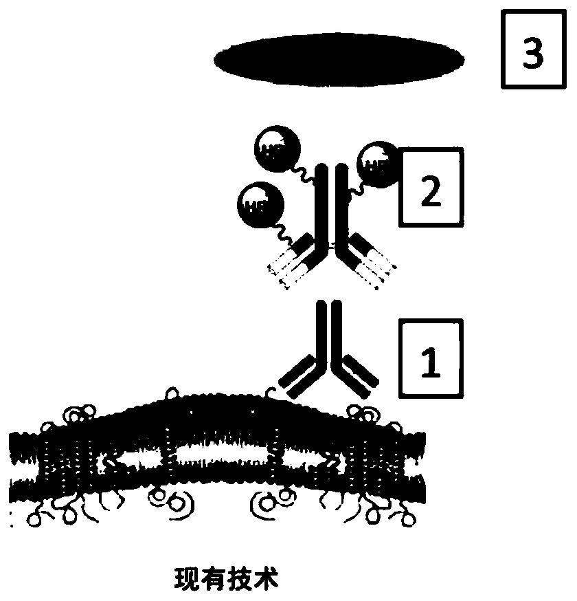
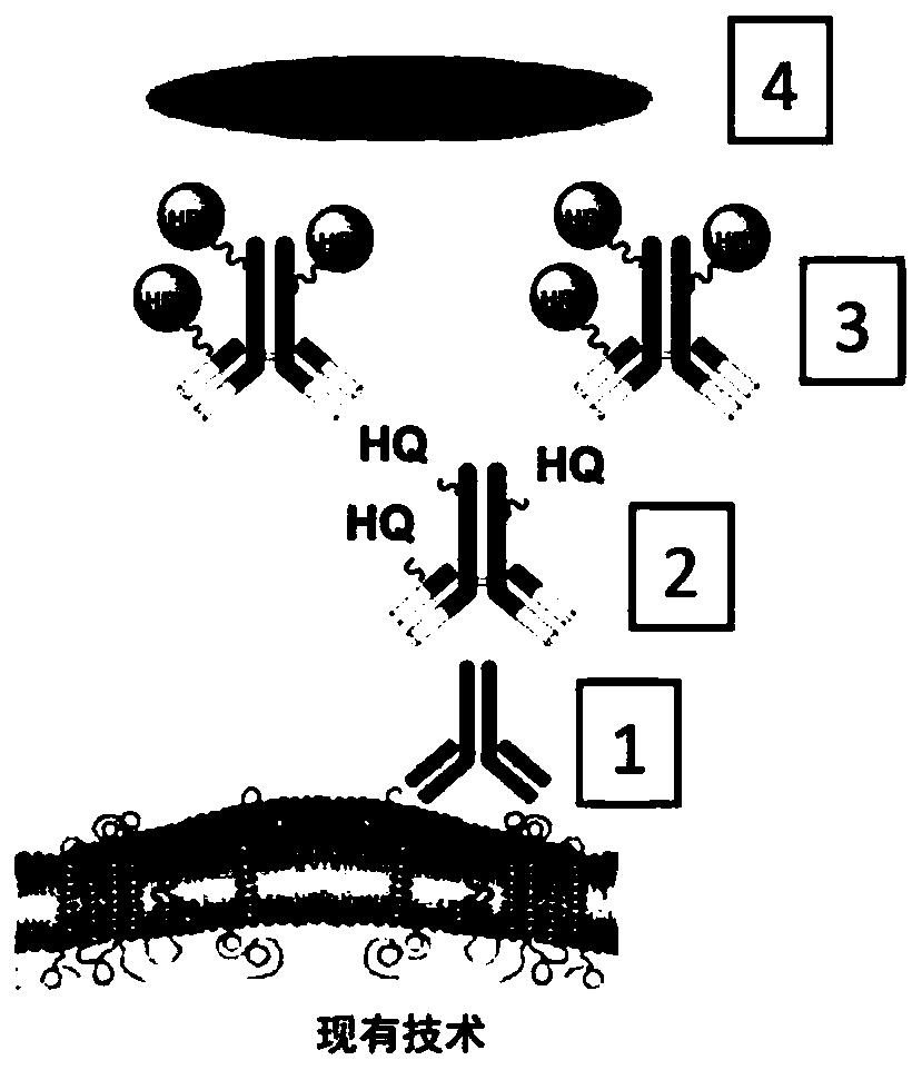
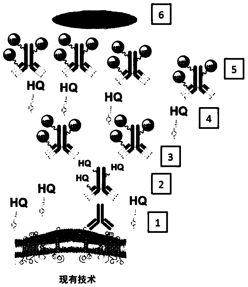
Methods and systems are provided for quantitative immunohistochemistry (IHC) of a target protein molecule including a secreted target protein molecule. The method comprises introducing to the sample:a primary antibody specific for the target protein molecule; a secondary antibody conjugated to a secondary antibody enzyme, the secondary antibody is specific for the primary antibody; a tyramide conjugated with a tyramide hapten, wherein the secondary antibody enzyme catalyzes deposition of the tyramide hapten onto the sample; a tertiary antibody conjugated with a tertiary antibody enzyme, the tertiary antibody is specific for the tyramide hapten; and a chromogen, wherein the tertiary antibody enzyme catalyzes a reaction with the chromogen to make the chromogen visible. The chromogen is visible as a punctate dot using microscopy.

Description

[0001] field of invention [0002] The present invention relates to immunohistochemical techniques, and more particularly, methods and systems for quantitative immunohistochemistry. [0003] Background of the invention [0004] Current techniques used in the automated detection of prognostic and / or predictive protein biomarkers rely on a threshold number of biomarker molecules (eg, greater than 1, 1,000, 10,000, etc.) to generate a visible signal. Thus, the number of protein biomarker molecules required to generate a visible signal depends on the characteristics of the detection technology used, primarily sensitivity and specificity. As such, current techniques cannot detect or quantify individual protein molecules. As a result, almost all established clinical assessments of prognostic and / or predictive protein biomarkers are limited to the use of binary analysis: plus and minus, reflecting the presence or absence of a signal. [0005] The invention features a method of ampli...

Claims


Application Information

Patent Timeline
Patent Type & Authority Applications(China)
IPC IPC(8): G01N33/542G01N33/543G01N33/58C12Q1/28
CPCC12Q1/28G01N33/542G01N33/54306G01N33/581G01N33/5306G01N17/00G01N33/53
Inventor W·德Z(大卫)·江A·佩达塔
Owner VENTANA MEDICAL SYST INC
Learn More