
Accurate cell nucleus segmentation method

A cell nucleus and accurate technology, applied in image analysis, image data processing, instruments, etc., can solve problems such as the influence of cohesive organs, unclear boundaries of cell nuclei, and different shapes of cell nuclei, and achieve the effect of reducing the amount of calculation

Inactive Publication Date: 2019-09-27
UNIV OF ELECTRONICS SCI & TECH OF CHINA
View PDF4 Cites 9 Cited by
  • Summary
  • Abstract
  • Description
  • Claims
  • Application Information

AI Technical Summary

Problems solved by technology

[0009] For the segmentation of cell nuclei similar to glioma, the present invention proposes an accurate segmentation method for cell nuclei, which can solve the problems of unclear boundaries of nuclei, different shapes of nuclei, and the influence of adhesion organs in the segmentation of cell nuclei similar to glioma. Nuclei are accurately segmented

Method used


Image

  • Accurate cell nucleus segmentation method
  • Accurate cell nucleus segmentation method
  • Accurate cell nucleus segmentation method

Examples


Embodiment Construction

[0058] The technical solution of the present invention will be described in detail below in conjunction with the accompanying drawings and specific embodiments.

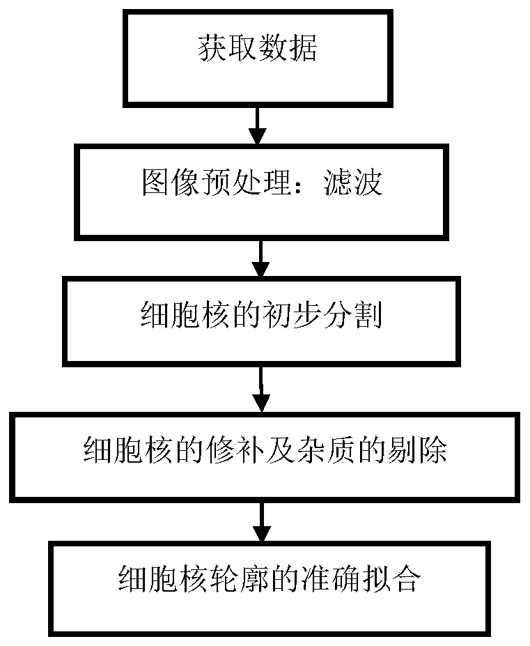
[0059] The present invention realizes the clinical segmentation of glioma cell nuclei, including data acquisition, image preprocessing steps, preliminary segmentation of cell nuclei, accurate fitting of cell nucleus contours, and segmentation of cohesive cell nuclei. The specific steps are as follows: figure 1 shown.

[0060] Step 1) Get data. The data comes from clinical images, which are private images made by sectioning human tissue and imaged with a slide scanner.

[0061] In this embodiment, glioma cell nuclei are taken as an example, and it is worth noting that the present invention can also be used to segment other cell nuclei of the same type. All the data in step 1) were provided by the Department of Pathology and Imaging of Southwest Hospital, and these data sets were collected from patients with glioma. ...


PUM


Abstract

An accurate cell nucleus segmentation method comprises the following steps: firstly, obtaining image data containing cell nuclei, and then carrying out pre-processing filtering operation on an image; carrying out preliminary segmentation on the cell nucleus by using a binary clustering algorithm; the influence of randomness of initial clustering selection on a segmentation result is reduced; selecting specific areas containing respective colors at the target and the background to iteratively update respective clustering centers, classifying all pixel points in the filtered image by using the obtained target clustering center and background clustering center as standards, judging whether each pixel point belongs to a target class or a background class, and finishing preliminary cell nucleus segmentation; repairing the preliminarily segmented cell nuclei, removing impurities, and performing accurate contour fitting on the cell nuclei by adopting a level set method; carrying out blocking operation of a partial overlapping area on the cell nucleus image subjected to preliminary segmentation, repairing and impurity removal, carrying out level set iteration on each block, and restoring the image according to a result to obtain an accurately segmented cell nucleus image.

Description

technical field [0001] The invention belongs to the field of computer-aided diagnosis and the field of medical clinical application, and relates to an accurate segmentation method of cell nuclei, which is of great significance for assisting doctors in diagnosing diseases, and improves the efficiency of doctors while ensuring the accuracy of segmentation. Background technique [0002] Changes in people's living environment and lifestyle have led to an increase in the incidence of cancer diseases affected by genetic and environmental factors. Especially in the past ten years, the incidence of cancer has been increasing year by year, and the mortality caused by cancer has become one of the important causes of death in China in recent years. Gliomas are malignant lesions of the glia in the brain and spinal cord. According to investigations, the incidence rate of this disease can reach 3-8 people / 100,000 population. This disease is caused by genetic factors on the one hand, and...

Claims


Application Information

Patent Timeline
IPC IPC(8): G06T7/00G06T7/11G06T7/13G06T7/136G06T7/90
CPCG06T7/0012G06T7/11G06T7/13G06T7/136G06T7/90G06T2207/30024G06T2207/30096
Inventor 李纯明董丹茹
Owner UNIV OF ELECTRONICS SCI & TECH OF CHINA
Learn More