Electric excision biopsy device for sheath of fiberoptic ductoscopy

A technique of excision biopsy and ductoscopy, applied in the field of medical devices, can solve the problems of low success rate and inability to be widely carried out, and achieve the effect of convenient accommodation

Pending Publication Date: 2019-08-20
崔建春
View PDF0 Cites 0 Cited by
  • Summary
  • Abstract
  • Description
  • Claims
  • Application Information

AI Technical Summary

Problems solved by technology

This method is only suitable for pedunculated lesions, and because the pedicle of space-occupying lesions is...

Method used


Image

  • Electric excision biopsy device for sheath of fiberoptic ductoscopy
  • Electric excision biopsy device for sheath of fiberoptic ductoscopy
  • Electric excision biopsy device for sheath of fiberoptic ductoscopy

Examples


Embodiment 1

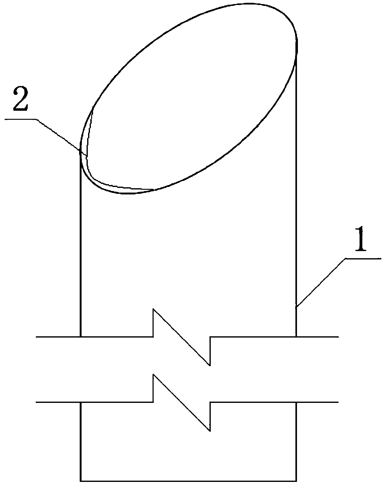
[0025] Such as Figure 1-3 As shown, a mammary ductoscope endoscopic electrosurgical excision biopsy device is composed of an endoscopic sheath 1 and an electrotome 2, wherein:

[0026] The mirror sheath 1 is a tubular structure with a circular or elliptical cross-section, and its front end is a sloped structure.

[0027] The electric knife 2 is arc-shaped, and its two ends are connected with the mirror sheath 1. The electric knife 2 is located on the lower side of the front slope of the mirror sheath 1; when in use, the low point of the mirror sheath slope is slightly higher than the level of the milk tube lens Location.

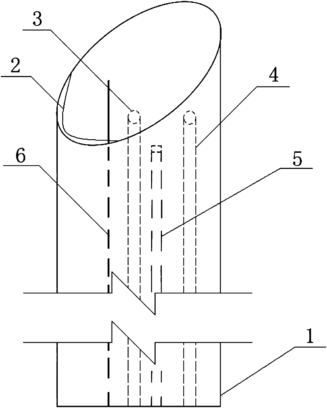
[0028] The electric knife 2 is connected to the wire, and the wire is placed inside the wall of the mirror sheath 1 to run. When it is near the tail of the mirror sheath 1, it is separated from the mirror sheath 1 and connected to the external power supply.

[0029] The wall of the mirror sheath 1 is also provided with a flushing channel 3 and a suction c...

Embodiment 2

[0034] The difference with Example 1 is:

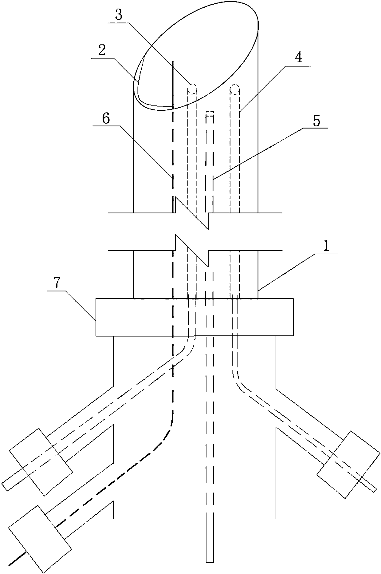
[0035] Such as Figure 4 As shown, there is no irrigation channel 3 and suction channel 4 on the inner wall of the mirror sheath 1, but the irrigation catheter 8 and the suction catheter 9 are used to realize the corresponding functions. The irrigation catheter 8 and the suction catheter 9 pass through the multi-channel adapter 7 (three channels) Insert into the mirror sheath 1 separately or simultaneously to realize flushing and suction functions.

Embodiment 3

[0037] The difference with Example 1 is:

[0038] The flushing channel 3 and the suction channel 4 on the inner wall of the scope sheath 1 are combined into one channel. When flushing is required, the flushing catheter is inserted into the channel through the multi-channel adapter for flushing. When suction is required, the flushing catheter is pulled out and the suction The catheter is inserted into the channel through a multi-channel conversion joint (three channels) for suction, so as to realize the flushing and suction functions.


PUM


Abstract

The invention aims to provide an electric excision biopsy device for a sheath of a fiberoptic ductoscopy, so as to excise a space occupying lesion in a laticifer with an electric knife. The electric excision biopsy device is characterized in that the electric excision biopsy device consists of the sheath and the electric knife, wherein the sheath adopts a pipe-shaped structure of which the sectionis circular or elliptic, and the front end of the sheath adopts a bevel structure; the electric knife penetrates across the bevel structure at the front end of the sheath or is in arc-shaped coincidence with a low-point position of the bevel structure; two ends of the electric knife are connected with the sheath; the electric knife is located on one side in a lower position of the bevel at the front end of the sheath; and the electric knife is connected with a sheath external power source through a conducting wire.

Description

technical field [0001] The invention relates to the field of medical equipment, and in particular provides a mammary ductoscope mirror sheath electric excision biopsy device. Background technique [0002] Ductoscopy has been routinely used in the diagnosis of ductal lesions, but ductoscopy can only detect space-occupying lesions in ductoscopy. There is no effective equipment and method for accurate excision and biopsy under ductoscopy. [0003] During the operation of ductoscopy, common accessories include: scope sheath, adapter, positioning hook, biopsy basket, etc. [0004] Previously, the existing ductoscopic excisional biopsy methods include: [0005] 1. Open a window through the side wall of the front end of the mirror sheath, the window is sharp, place the space-occupying lesion in the milk duct into the window of the side wall of the mirror sheath, and use the forward and backward movement of the mirror sheath to remove the lesion in the milk duct. The main disadvan...

Claims


Application Information

Patent Timeline
IPC IPC(8): A61B18/14A61B10/04
CPCA61B18/14A61B10/04A61B2018/1405A61B2018/00982A61B2218/002A61B2218/007
Inventor 崔建春裴晓华张董晓桂冰陈凯吴雪卿李立柳青峰董齐高洋马帅徐洋
Owner 崔建春
Why Eureka
Try Eureka