
Medical image recognition system and method based on multi-modal fusion

A medical image and recognition system technology, applied in the field of medical image detection, can solve the problems of inability to integrate various medical images, low recognition accuracy, poor detection comprehensiveness, etc., to improve recognition stability and accuracy, and high recognition accuracy , The effect of improving the generalization ability

Active Publication Date: 2019-06-07
青岛中科智康医疗科技有限公司
View PDF7 Cites 8 Cited by
  • Summary
  • Abstract
  • Description
  • Claims
  • Application Information

AI Technical Summary

Problems solved by technology

[0003] The purpose of the present invention is: the present invention provides a medical image recognition system and method based on multi-modal fusion, which solves the problem that various medical images cannot be integrated due to inconsistency of medical image characteristics, poor comprehensiveness of detection, and low recognition accuracy The problem

Method used


Image

  • Medical image recognition system and method based on multi-modal fusion
  • Medical image recognition system and method based on multi-modal fusion
  • Medical image recognition system and method based on multi-modal fusion

Examples


Embodiment 1

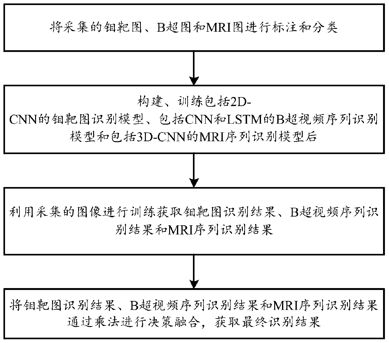
[0091] A medical image recognition system based on multimodal fusion, including

[0092] Mammogram recognition model, used to construct 2D-CNN to obtain mammogram features and complete mammogram recognition;

[0093] B-ultrasound video sequence recognition model, used to construct CNN and LSTM to obtain B-ultrasound video sequence features, and complete B-ultrasound video sequence recognition;

[0094] MRI sequence recognition model, used to construct 3D-CNN to obtain MRI sequence features and complete MRI sequence recognition;

[0095] The multimodal decision-making fusion unit is used for decision-making fusion of mammogram recognition results, B-ultrasound video sequence recognition results and MRI sequence recognition results through multiplication to obtain the final recognition result.

[0096] A medical image recognition method based on multimodal fusion, comprising the steps of:

[0097] Step 1: Label and classify the collected mammograms, B-ultrasound images and MRI...

Embodiment 2

[0102] Based on embodiment 1, first construct the recognition network of corresponding image, and it is trained to obtain mammogram recognition model, B supersonic video sequence recognition model and MRI sequence recognition model;

[0103] In step 2, the mammogram recognition model training includes the following steps:

[0104] Step a1: Build a mammogram recognition model, which includes sequentially connected 2D-CNN, fully connected layer and Softmax;

[0105] Step a2: Input the training set in the step 1 classified image into the mammogram recognition model to train at intervals, and record the model training parameters until the objective function curve no longer declines;

[0106] Step a3: Input the verification set in the classification image into the mammogram recognition model in step a2 for testing, and record six models with good recognition effect;

[0107] Step a4: Input the test set data into the six models mentioned in step a3, and select the one with the high...


PUM


Abstract

The invention discloses a medical image recognition system and method based on multi-modal fusion, and relates to the field of medical image detection. The system comprises a molybdenum target image recognition model which is used for constructing a 2D-CNN structure to obtain molybdenum target image characteristics to complete molybdenum target image recognition, a B-mode ultrasound video sequencerecognition model which is used for constructing a CNN and LSTM structure to obtain B-mode ultrasound sequence characteristics to complete B-mode ultrasound video sequence recognition, and an MRI sequence recognition model which is used for constructing a 3D-CNN to obtain MRI sequence characteristics to complete MRI sequence recognition, and a multi-modal decision fusion unit which is used for carrying out decision fusion on molybdenum target image recognition result, B-mode ultrasound video sequence recognition result and MRI sequence recognition result through multiplication to obtain finalrecognition result. According to the method, different structures are designed for training according to the characteristics of the molybdenum target, the B-ultrasonic image and the MRI image, the difficulty of combining the images with completely inconsistent characteristics is overcome, decision fusion is performed on multi-dimensional recognition results through multiplication, more comprehensive and effective characteristics are obtained, and the recognition accuracy is improved.

Description

technical field [0001] The invention relates to the field of medical image detection, in particular to a medical image recognition system and method based on multimodal fusion. Background technique [0002] At present, medical imaging detection is the preferred method to determine breast cancer and other diseases, and has achieved certain results. At present, hospitals mainly conduct B-ultrasound, mammography, and MRI detection for breast cancer, and manually analyze and process the detection images. Since the density of soft tissues such as glands, blood vessels, and fat in breast tissue is very close to the density of the lesion area, it is more difficult to observe with the naked eye. Visual fatigue is easy to cause misdiagnosis; with the continuous development of medical imaging technology and computer technology, it is possible to use computer to assist diagnosis; Features, use SVM and other machine learning methods to classify and identify the parameters of breast dis...

Claims


Application Information

Patent Timeline
IPC IPC(8): G06K9/62G06N3/04
CPCY02T10/40
Inventor 韩云翟红波
Owner 青岛中科智康医疗科技有限公司
Learn More