Calcified cluster positioning method and device, computer equipment and storage medium

A positioning method and calcification point technology, applied in the field of medical devices, can solve the problems of complex methods, affecting the speed and accuracy of positioning, and large amount of calculation data.

Active Publication Date: 2019-05-24
SHANGHAI UNITED IMAGING HEALTHCARE
View PDF7 Cites 5 Cited by
  • Summary
  • Abstract
  • Description
  • Claims
  • Application Information

AI Technical Summary

Problems solved by technology

[0003] Traditional detection methods for calcified areas in breast three-dimensional tomographic images, when processing, some need to use the scanned original image, combined with the original image and the reconstructed image for detection, some are processed on a single image, and finally Then synthesize it into 3D images, this method is more complicated, and the amount of calculation data is large, which affects the speed and accuracy of positioning

Method used


Image

  • Calcified cluster positioning method and device, computer equipment and storage medium
  • Calcified cluster positioning method and device, computer equipment and storage medium
  • Calcified cluster positioning method and device, computer equipment and storage medium

Examples


Embodiment Construction

[0050] In order to make the purpose, technical solution and advantages of the present application clearer, the present application will be further described in detail below in conjunction with the accompanying drawings and embodiments. It should be understood that the specific embodiments described here are only used to explain the present application, and are not intended to limit the present application.

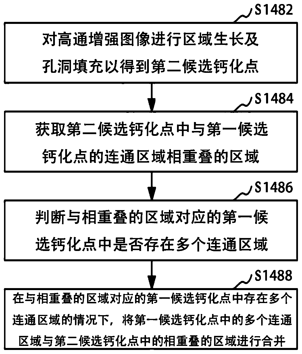
[0051] figure 1 It is a schematic flow chart of a method for locating calcification clusters in an embodiment, such as figure 1 As shown, a method for locating calcification clusters is applied to breast three-dimensional tomographic images, including:

[0052] Step S120: Obtain a space of interest in the three-dimensional tomographic image of the breast.

[0053] Specifically, when detecting and locating calcification clusters, firstly, a digital breast tomography (Digital Breast Tomsynthesis, referred to as DBT) technique can be used, that is, a fixed breast is exposed...


PUM


Abstract

The invention relates to a calcified cluster positioning method and device, computer equipment and a storage medium, and the calcified cluster positioning method is applied to a mammary gland three-dimensional tomographic image, and comprises the steps: obtaining an interested space in the mammary gland three-dimensional tomographic image; segmenting the space of interest to obtain candidate calcification points; screening out and determining a calcification point from the candidate calcification points; and clustering the determined calcification points to obtain a calcification cluster region. According to the positioning method of the calcified cluster, the interested space is selected from the mammary gland three-dimensional tomographic image; According to the method, the interested space can be segmented and screened, the calcification points are clustered after being obtained, so that positioning of the calcification clusters is achieved, only reconstructed mammary gland three-dimensional tomography image data are needed in the positioning process, scanned original data are not needed, the processed data size is reduced, and positioning of the calcification clusters is rapidand accurate.

Description

technical field [0001] The invention relates to the field of medical devices, in particular to a method, device, computer equipment and storage medium for locating calcification clusters. Background technique [0002] Three-dimensional breast tomography (Digital Breast Tomsynthesis, referred to as DBT) can significantly reduce the overlap of breast tissue, and can better observe breast tissue lesions. Locating the calcified areas that are only present in a portion of the image can aid in faster interpretation. [0003] Traditional detection methods for calcified areas in breast three-dimensional tomographic images, when processing, some need to use the scanned original image, combined with the original image and the reconstructed image for detection, some are processed on a single image, and finally And then synthesized into 3D images, such a method is relatively complicated, and the amount of calculation data is large, which affects the speed and accuracy of positioning. ...

Claims


Application Information

Patent Timeline
Patent Type & Authority Applications(China)
IPC IPC(8): G06T7/00G06T7/11G06T7/70G06T17/00G06T5/00
Inventor 吴叶芬
Owner SHANGHAI UNITED IMAGING HEALTHCARE
Why Eureka
Try Eureka