
X-ray-imaging automatic exposure control method, storage medium and medical equipment

A technology of automatic exposure and control method, applied in the field of medical devices, can solve the problems of increasing the cost of the detector, increasing the complexity of the detector, etc., and achieve the effect of flexible and diverse ROI range, reducing the requirements of positioning accuracy, and high-performance system

Active Publication Date: 2019-05-14
SIEMENS SHANGHAI MEDICAL EQUIP LTD
View PDF9 Cites 10 Cited by
  • Summary
  • Abstract
  • Description
  • Claims
  • Application Information

AI Technical Summary

Problems solved by technology

This increases the complexity of the detector and thus increases the cost of the detector

Method used


Image

  • X-ray-imaging automatic exposure control method, storage medium and medical equipment
  • X-ray-imaging automatic exposure control method, storage medium and medical equipment
  • X-ray-imaging automatic exposure control method, storage medium and medical equipment

Examples


Embodiment Construction

[0045] In order to have a clearer understanding of the technical features, purposes and effects of the present invention, the specific implementation manners of the present invention will now be described with reference to the accompanying drawings, in which the same symbols represent the same parts.

[0046] In this article, "schematic" means "serving as an example, example or illustration", and any illustration or implementation described as "schematic" should not be interpreted as a more preferred or more advantageous Technical solutions.

[0047]In order to make the drawing concise, each drawing only schematically shows the parts related to the present invention, and they do not represent the actual structure of the product. In addition, to make the drawings concise and easy to understand, in some drawings, only one of the components having the same structure or function is schematically shown, or only one of them is marked.

[0048] In this article, "one" and "one" not o...


PUM


Abstract

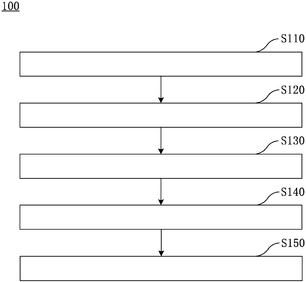
The invention relates to an X-ray-imaging automatic exposure control method, a storage medium and medical equipment. According to one execution mode, the X-ray-imaging automatic exposure control method includes the steps that a to-be-detected object is pre-exposed with the set pre-exposure dose Mp, and a low-dose exposure image is obtained; the outline of the to-be-detected object is determined onthe low-dose exposure image; on the low-dose exposure image, based on a selected pixel position, an interested region ROI is defined in the outline range of the to-be-detected object; the average pixel value G<ROI> of the interested region ROI is calculated; according to the average pixel value G<ROI>of the interested region ROI, the main exposure dose Mr is calculated. According to the X-ray-imaging automatic exposure control method, the storage medium and the medical equipment, a physical AEC chamber does not need to be used, and the requirement for the placement accuracy is reduced while the image quality is guaranteed.

Description

technical field [0001] The invention relates to the technical field of medical equipment, in particular to an automatic exposure control method for X-ray imaging, a computer storage medium and X-ray medical equipment. Background technique [0002] Usually, an AEC room is set in front of the detector of the X-ray medical equipment to calculate the estimated dose of ROI (Region of Interest) for each inspection. The more AEC chambers, the higher the accuracy of measurement, but this leads to an increase in cost. [0003] Several solutions have been proposed that do not use an AEC chamber, some of which involve utilizing some pixels or groups of pixels to function as AEC chambers. These pixels or groups of pixels are controlled and read one by one. As such, the complexity and thus the cost of the detector is increased. For example, the Chinese invention patent application with the publication number CN106550527A relates to a method of acquiring X-ray images by arranging AEC p...

Claims


Application Information

Patent Timeline
IPC IPC(8): A61B6/00
Inventor 赫伟
Owner SIEMENS SHANGHAI MEDICAL EQUIP LTD
Learn More