
Medical image abnormal region segmentation method and device

A technology of medical images and abnormal areas, applied in the field of medical image processing, can solve the problems of poor accuracy and achieve the effect of improving accuracy and recognition efficiency

Pending Publication Date: 2019-03-19
SHANGHAI EAGLEVISION MEDICAL TECH CO LTD
View PDF5 Cites 24 Cited by
  • Summary
  • Abstract
  • Description
  • Claims
  • Application Information

AI Technical Summary

Problems solved by technology

The existing technology uses machine learning models such as convolutional neural networks to identify medical images, and the accuracy of finding small abnormal areas is relatively poor

Method used


Image

  • Medical image abnormal region segmentation method and device
  • Medical image abnormal region segmentation method and device
  • Medical image abnormal region segmentation method and device

Examples


Embodiment Construction

[0037] The technical solutions of the present invention will be clearly and completely described below in conjunction with the accompanying drawings. Apparently, the described embodiments are some of the embodiments of the present invention, but not all of them. Based on the embodiments of the present invention, all other embodiments obtained by persons of ordinary skill in the art without making creative efforts belong to the protection scope of the present invention.

[0038] In the description of the present invention, it should be noted that the terms "center", "upper", "lower", "left", "right", "vertical", "horizontal", "inner", "outer" etc. The indicated orientation or positional relationship is based on the orientation or positional relationship shown in the drawings, and is only for the convenience of describing the present invention and simplifying the description, rather than indicating or implying that the referred device or element must have a specific orientation, ...


PUM


Abstract

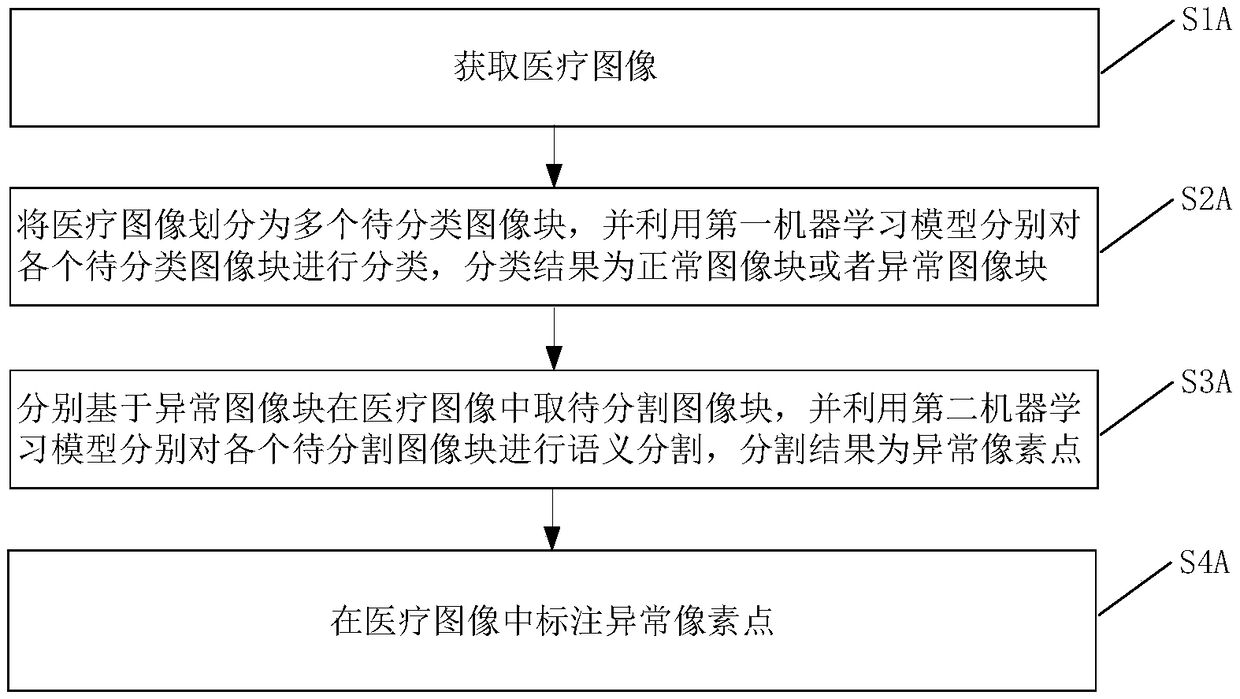
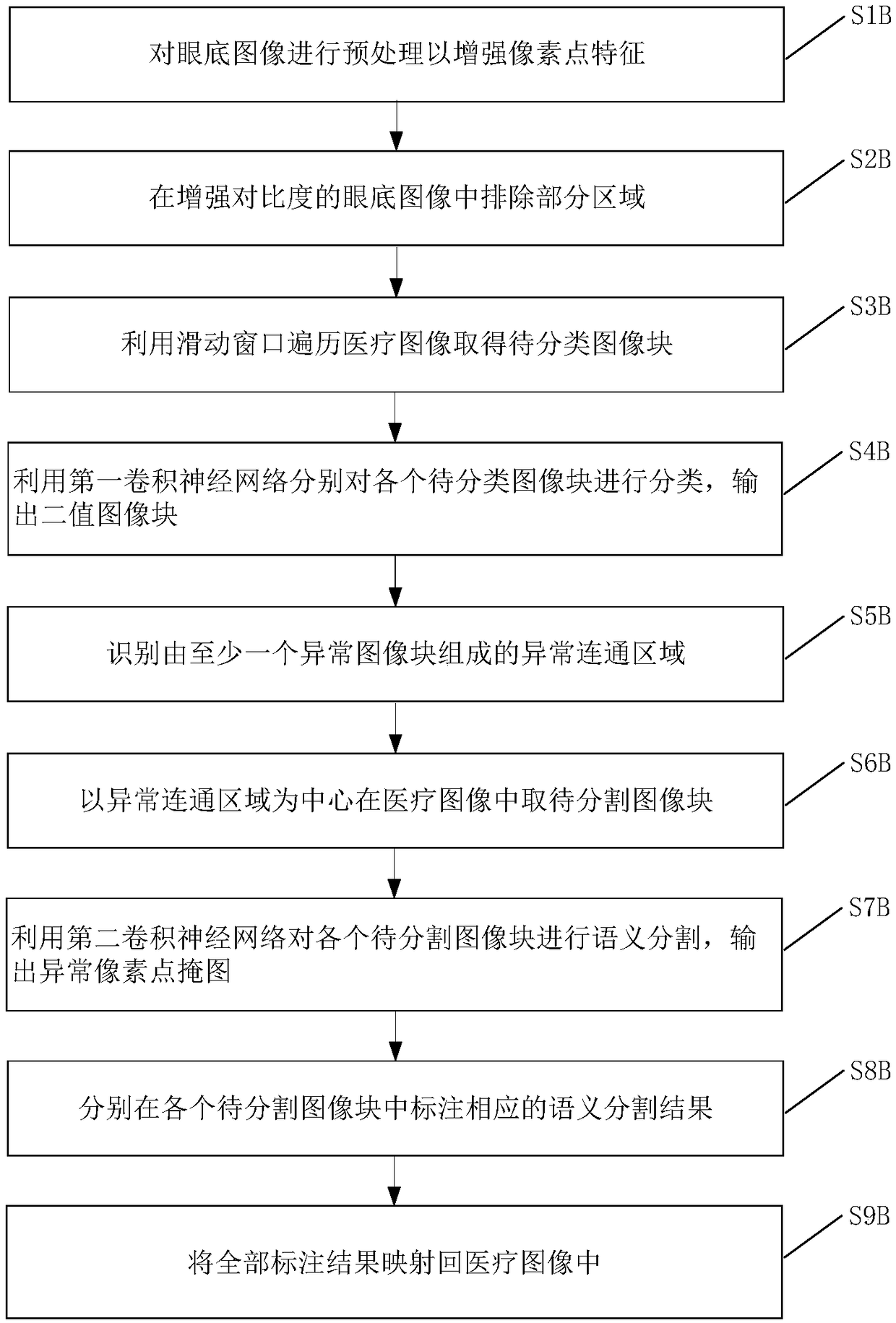
The invention provides a medical image abnormal region segmentation method and device. The method comprises the steps of obtaining a medical image; dividing the medical image into a plurality of to-be-classified image blocks, and utilizing a first machine learning model to classify each to-be-classified image block, the classification result being a normal image block or an abnormal image block; respectively taking to-be-segmented image blocks from the medical image based on the abnormal image blocks, and respectively performing semantic segmentation on each to-be-segmented image block by using a second machine learning model, the segmentation result being an abnormal pixel point; and marking the abnormal pixel points in the medical image.

Description

technical field [0001] The invention relates to the field of medical image processing, in particular to a method and equipment for segmenting abnormal regions of medical images. Background technique [0002] In the field of computer vision, the current hot research directions mainly include image recognition, target location and detection, semantic segmentation, etc. Image segmentation is an essential part of computer vision and a key step from image processing to image analysis. [0003] Image segmentation refers to dividing an image into several disjoint regions based on features such as gray scale, color, spatial texture, geometric shape, etc., so that these features show consistency or similarity in the same region, but appear in different regions. obvious difference. Semantic segmentation is to identify the semantics of each area on the basis of image segmentation. Image semantic segmentation is an important branch in the field of AI, and is currently widely used in ...

Claims


Application Information

Patent Timeline
IPC IPC(8): G06T7/00G06T7/11G06T7/187
CPCG06T7/0012G06T2207/10081G06T2207/10132G06T2207/20081G06T2207/30041G06T2207/30096G06T7/11G06T7/187
Inventor 黄烨霖熊健皓赵昕张大磊
Owner SHANGHAI EAGLEVISION MEDICAL TECH CO LTD
Learn More