
A Brain Tissue Segmentation Method Based on Regularized Graph Cut

A technology of brain tissue and tissue, applied in brain magnetic resonance image processing, brain tissue segmentation based on regularized graph cuts, can solve problems such as high computational complexity, and achieve the effect of suppressing noise, good segmentation effect, and suppressing influence

Active Publication Date: 2021-11-02
SOUTHEAST UNIV
View PDF3 Cites 0 Cited by
  • Summary
  • Abstract
  • Description
  • Claims
  • Application Information

AI Technical Summary

Problems solved by technology

The voxel segmentation method can achieve excellent performance, but in MRI, when facing tens of thousands of supervoxels, it will encounter high computational complexity, which limits its application in MRI

Method used


Image

  • A Brain Tissue Segmentation Method Based on Regularized Graph Cut
  • A Brain Tissue Segmentation Method Based on Regularized Graph Cut
  • A Brain Tissue Segmentation Method Based on Regularized Graph Cut

Examples


Embodiment Construction

[0046] The technical solutions provided by the present invention will be described in detail below in conjunction with specific examples. It should be understood that the following specific embodiments are only used to illustrate the present invention and are not intended to limit the scope of the present invention.

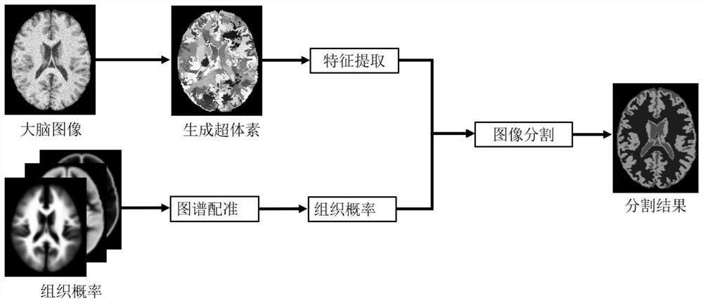
[0047] A brain tissue segmentation method based on regularized graph cut provided by the present invention, the process is mainly divided into two parts: first, using the super-voxel generation method based on intensity distance and spatial similarity, the voxels are divided into regular, To reduce the influence of noise, it can better fit the supervoxels in the edge area of ​​the image; then, combined with the prior knowledge of brain tissue, use graph cut to cut each supervoxel into specific brain tissue. Concrete flow process of the present invention is as figure 1 shown, including the following steps:

[0048] Step 1: Generation of supervoxels:

[0049]The ...


PUM


Abstract

The invention discloses a method for brain tissue segmentation based on regularized graph cuts. First, based on intensity distance and spatial similarity, a new calculation method for similarity between voxels is designed, so as to cluster voxels and segment brain MRI images. It is a series of supervoxels that evenly fit the edge of the image; then Ming designs an energy calculation formula by integrating the prior probability of different tissues of the brain into the graph cut framework, and calculates the distribution of each supervoxel in different The label is the energy value of each part, so the graph cut method is used to segment the supervoxel, and the Magnetic Resonance Imaging (MRI) image is divided into different tissues. The present invention can segment three kinds of brain tissues from the initial brain MRI, and in the segmentation result, the boundaries between the various tissues have a high degree of fit. Compared with the existing MRI image segmentation method, the invention has better segmentation effect, higher boundary fitting degree, higher efficiency, faster processing speed, and can better suppress the influence of noise.

Description

technical field [0001] The invention belongs to the technical field of digital image processing, and relates to a brain magnetic resonance image processing method, more specifically, to a brain tissue segmentation method based on a regularized graph cut. Background technique [0002] Magnetic resonance imaging (MRI) has been widely used to examine the anatomy of the human brain in clinical applications and neuroscience research. Compared with other medical imaging modalities, MRI has the advantages of high spatial resolution and good soft tissue contrast, which can finely distinguish different types of tissues. Accurate segmentation of these tissues is critical for some applications. [0003] In order to facilitate the processing of MRI images, the concept of supervoxel is introduced. Supervoxel (supervoxel) or superpixel (superpixel) is an image preprocessing technology that has developed rapidly in recent years. Compared with the basic unit in traditional processing met...

Claims


Application Information

Patent Timeline
Patent Type & Authority Patents(China)
IPC IPC(8): G06T7/162G06T7/11
CPCG06T2207/10088G06T2207/20072G06T2207/30016G06T7/11G06T7/162
Inventor 章品正李艺飞孔佑勇伍家松杨淳沨舒华忠
Owner SOUTHEAST UNIV
Learn More