Quantitative myocardial magnetic resonance imaging method and equipment and storage medium

A magnetic resonance imaging and myocardial technology, applied in the field of medical imaging, can solve problems such as long scanning time, restricted imaging resolution, complex downsampling and reconstruction algorithms, etc., to achieve the effect of expanding imaging field of view and improving spatial resolution

Active Publication Date: 2018-12-25
TSINGHUA UNIV
View PDF6 Cites 3 Cited by
  • Summary
  • Abstract
  • Description
  • Claims
  • Application Information

AI Technical Summary

Problems solved by technology

The breath-hold requirement restricts further improvements in imaging resolution and cannot be used for subjects with difficulty in breath-holding (common in cardiac patients)
In the prior art, scanning acceleration is performed through parallel imaging technology, which sacrifices the signal-to-noise ratio of the signal, and introduces complex down-sampling and reconstruction algorithms
Finally, more T 1 and T 2 Only weighted sampling points can be imaged, so it is sensitive to heart rate changes and has a long scan time

Method used


Image

  • Quantitative myocardial magnetic resonance imaging method and equipment and storage medium
  • Quantitative myocardial magnetic resonance imaging method and equipment and storage medium
  • Quantitative myocardial magnetic resonance imaging method and equipment and storage medium

Examples


Embodiment Construction

[0031] In order to make the objects, technical solutions and advantages of the present invention more apparent, exemplary embodiments according to the present invention will be described in detail below with reference to the accompanying drawings. Apparently, the described embodiments are only some embodiments of the present invention, rather than all embodiments of the present invention, and it should be understood that the present invention is not limited by the exemplary embodiments described here. Based on the embodiments of the present invention described in the present invention, all other embodiments obtained by those skilled in the art without creative effort shall fall within the protection scope of the present invention.

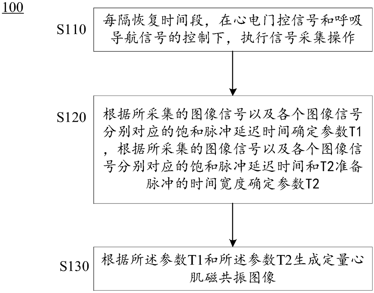
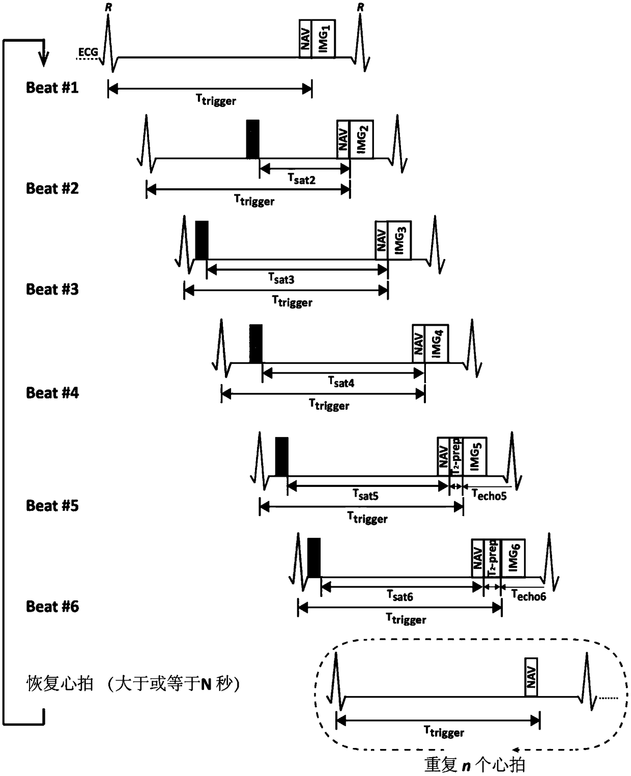
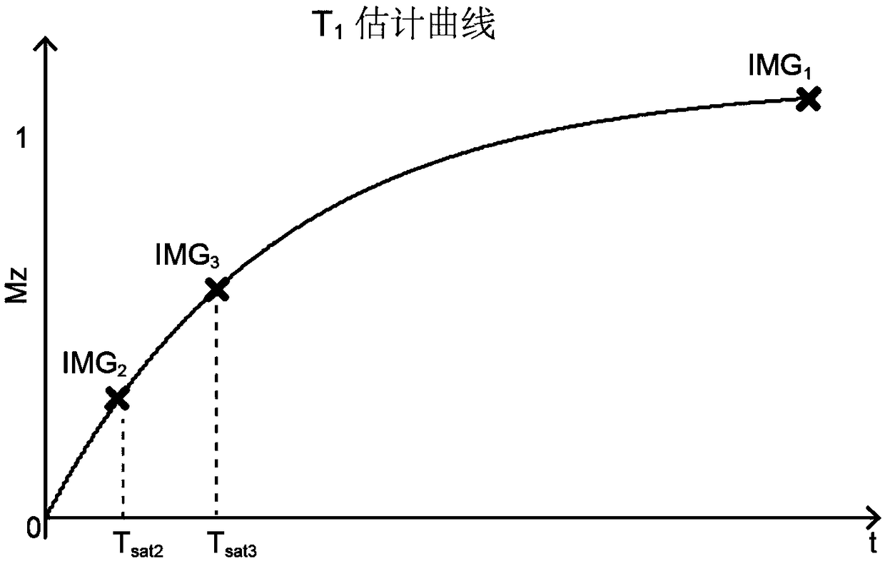
[0032] According to an embodiment of the present invention, a quantitative myocardial magnetic resonance imaging method is provided. The method is a 3D free-breathing quantitative myocardial parameter T 1 and T 2 Combined Imaging Technology. The...


PUM


Abstract

The invention provides a quantitative myocardial magnetic resonance imaging method and equipment and a storage medium. The method comprises the steps that image signal acquisition operation is performed under the control of an electrocardiograph gating signal and respiratory navigation signal every recovery time period; a parameter T1 is determined according to an acquired image signal and delay time of a saturation pulse corresponding to the image signal, and a parameter T2 is determined according to the acquired image signal, the delay time of the saturation pulse corresponding to the imagesignal and an echo time interval of a T2 preparation pulse; a quantitative myocardial magnetic resonance image is generated according to the parameter T1 and the T2. The quantitative myocardial magnetic resonance imaging method and equipment and the storage medium have the advantages that scanning can be completed under the condition that a testee breathes freely and does not need to suspend breathing; it is allowed that the imaging field is further enlarged and the spatial resolution is increased; in addition, completely stagger and segmental acquisition is performed among sampling points through k-space, thereby achieving the internal registration of an original image, and other image processing in a later stage is not needed.

Description

technical field [0001] The present invention relates to the field of medical imaging, and more specifically relates to a quantitative myocardial magnetic resonance imaging method, equipment and storage medium. Background technique [0002] Magnetic Resonance Imaging technology uses the phenomenon of nuclear magnetic resonance to image the human body, which is already a common medical imaging examination method. [0003] The basic physical parameter T of NMR 1 (spin-lattice relaxation time, or longitudinal relaxation time) and T 2 (Spin-spin relaxation time, or transverse relaxation time) is a time constant describing the recovery process of the longitudinal magnetization vector and the decay process of the transverse magnetization vector. T 1 and T 2 It is determined by the composition and structure of biological tissue and the strength of the magnetic field. Under a certain magnetic field strength, different tissues have specific T 1 and T 2 value. When biological t...

Claims


Application Information

Patent Timeline
Patent Type & Authority Applications(China)
IPC IPC(8): A61B5/055
CPCA61B5/0044A61B5/055A61B5/7203A61B5/7225A61B2576/023
Inventor 丁海艳郭瑞罗建文
Owner TSINGHUA UNIV
Why Eureka
Try Eureka