
Image recognition and analysis system and method of intestinal pathological section based on depth learning

A technology of pathological slices and image recognition, applied in the field of medical detection, can solve the problems of manual reading fatigue, affecting early diagnosis, treatment and prognosis of patients, and heavy workload, so as to help rational allocation and reduce the rate of missed diagnosis , the effect of high accuracy

Inactive Publication Date: 2018-12-21
WUHAN ENDOANGEL MEDICAL TECH CO LTD
View PDF4 Cites 6 Cited by
  • Summary
  • Abstract
  • Description
  • Claims
  • Application Information

AI Technical Summary

Benefits of technology

This technology uses advanced techniques from medicine (medicine) that can help diagnose diseases accurately by analyzing patient's medical image content or symptoms. It also includes machine learning tools used during analysis which improve its effectiveness over traditional methods while reducing errors caused by human error. Overall, these technical improvements make better ways to detect illnesses faster than current approaches such as histology examination.

Problems solved by technology

This patented technical problem addressed in this patents relates to improving the accuracy and efficiency of diagnosing illnesses caused by complex disease patterns through advanced techniques such as machine learning or computer vision analysis.

Method used


Image

  • Image recognition and analysis system and method of intestinal pathological section based on depth learning
  • Image recognition and analysis system and method of intestinal pathological section based on depth learning

Examples


Embodiment Construction

[0015] In order to facilitate those of ordinary skill in the art to understand and implement the present invention, the present invention will be described in further detail below in conjunction with the accompanying drawings and embodiments. It should be understood that the implementation examples described here are only used to illustrate and explain the present invention, and are not intended to limit this invention.

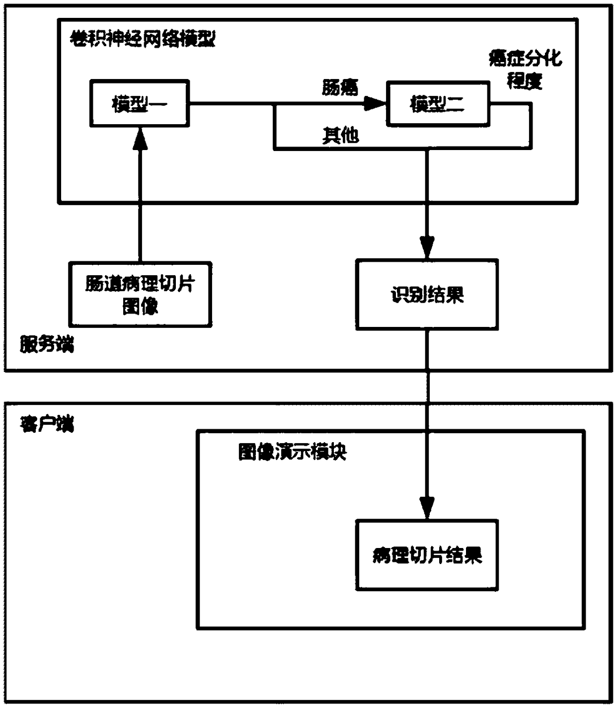
[0016] please see figure 1 , a deep learning-based intestinal pathological slice image recognition and analysis system provided by the present invention includes a client and a server; the client is used to monitor the collected intestinal pathological slice image and transmit it to the server, receive and display the service The analysis result fed back by the client; the server, according to the intestinal pathological slice image collected from the client, immediately judges the pathological result corresponding to the intestinal pathological slice image, ...


PUM


Abstract

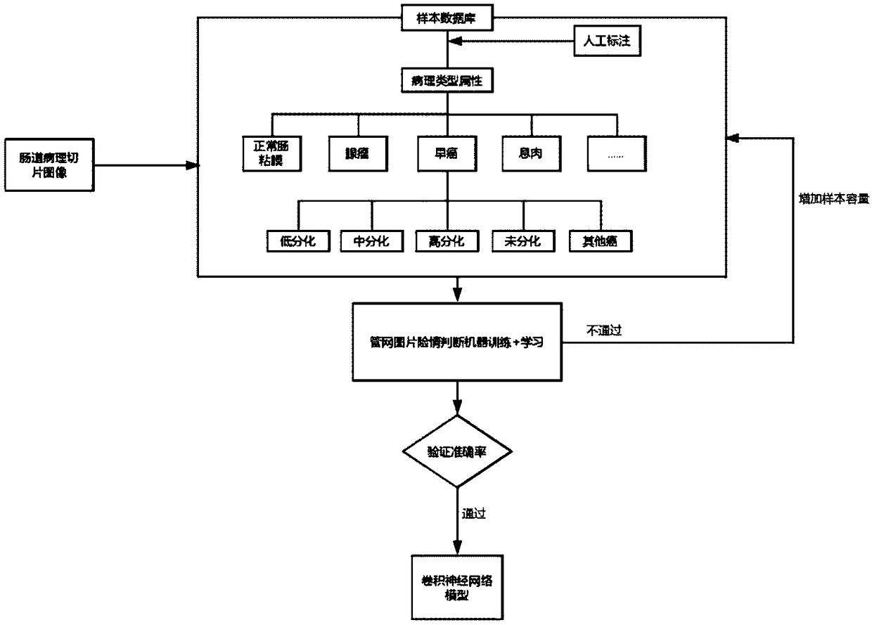
The invention discloses an intestinal pathological slice image recognition and analysis system and a method based on depth learning. The system comprises a client end and a server end. The client endis used for monitoring and transmitting the collected intestinal pathological slice images to the server end, receiving and displaying the analysis results fed back by the server end; The server judges the pathological result corresponding to the intestinal pathological slice image according to the intestinal pathological slice image collected from the client and feeds back the analysis result tothe client. At first, that client end collect and obtains the intestinal pathological slice image, and uploads the intestinal pathological slice image to the server end; Then, the server receives theintestinal pathological slice image as a parameter, calls the convolution neural network model for recognition, recognizes the features of the intestinal pathological slice image and outputs them. Finally, the client receives and displays the analysis results. The invention can provide accurate and reliable reference for physicians, improve the accuracy and effectiveness of pathological report, issimple and easy to use, and has remarkable social and economic value.

Description


Claims


Application Information

Patent Timeline
Owner WUHAN ENDOANGEL MEDICAL TECH CO LTD
Learn More