
Pancreatic cystic tumor CT image classification method based on multi-channel multiple classifiers

A multi-classifier, multi-channel technology, applied in image analysis, image enhancement, image data processing and other directions, can solve problems such as affecting results, artificial error classifiers, etc., to achieve the effect of improving accuracy, reducing errors, and enhancing edge information

Active Publication Date: 2018-11-27
ZHEJIANG UNIV OF TECH
View PDF6 Cites 8 Cited by
  • Summary
  • Abstract
  • Description
  • Claims
  • Application Information

AI Technical Summary

Problems solved by technology

[0004] In order to overcome the human error introduced by manual tumor segmentation and the insufficiency of a single classifier affecting the results

Method used


Image

  • Pancreatic cystic tumor CT image classification method based on multi-channel multiple classifiers
  • Pancreatic cystic tumor CT image classification method based on multi-channel multiple classifiers
  • Pancreatic cystic tumor CT image classification method based on multi-channel multiple classifiers

Examples


Embodiment Construction

[0046] The present invention will be further described below in conjunction with the accompanying drawings.

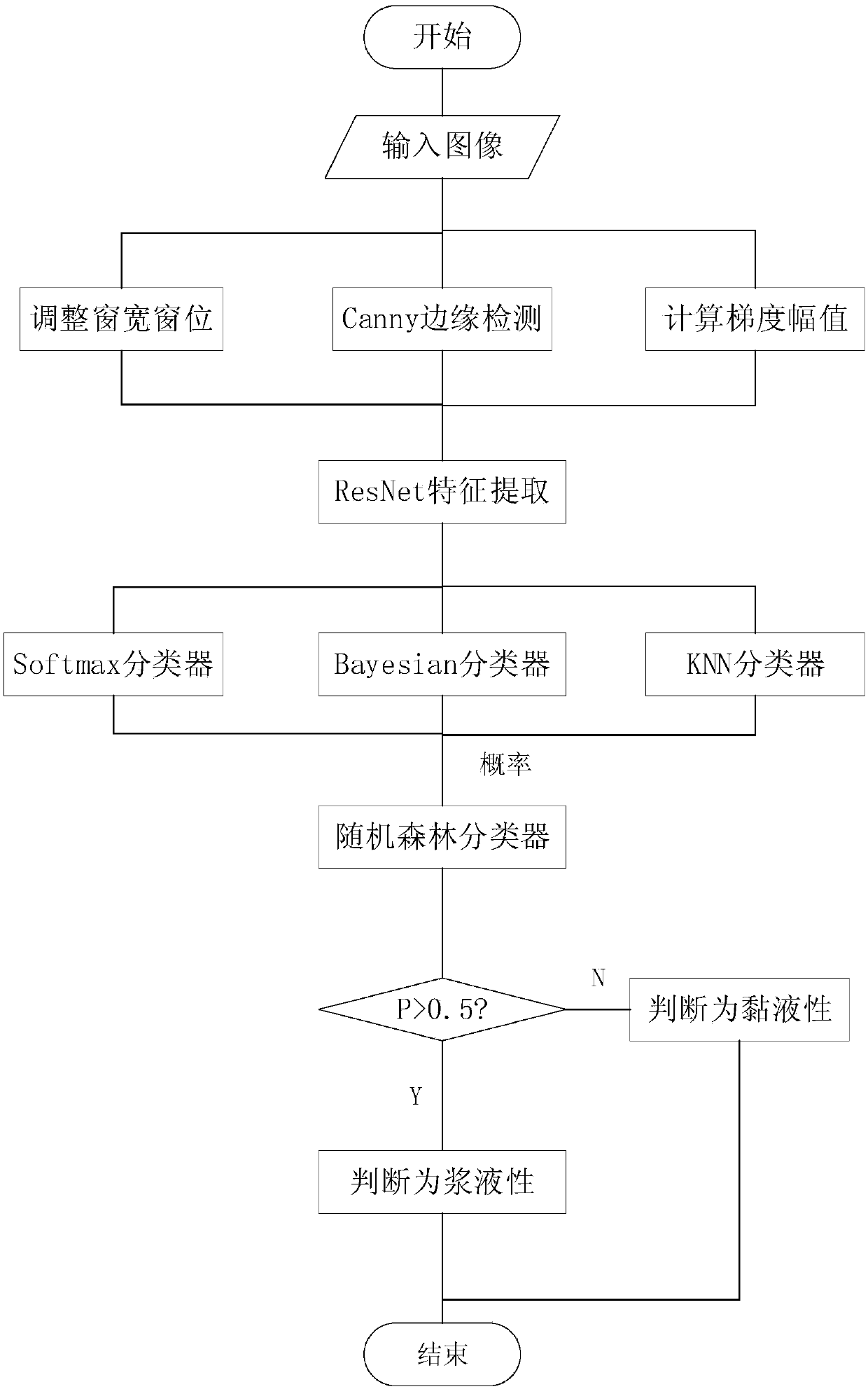
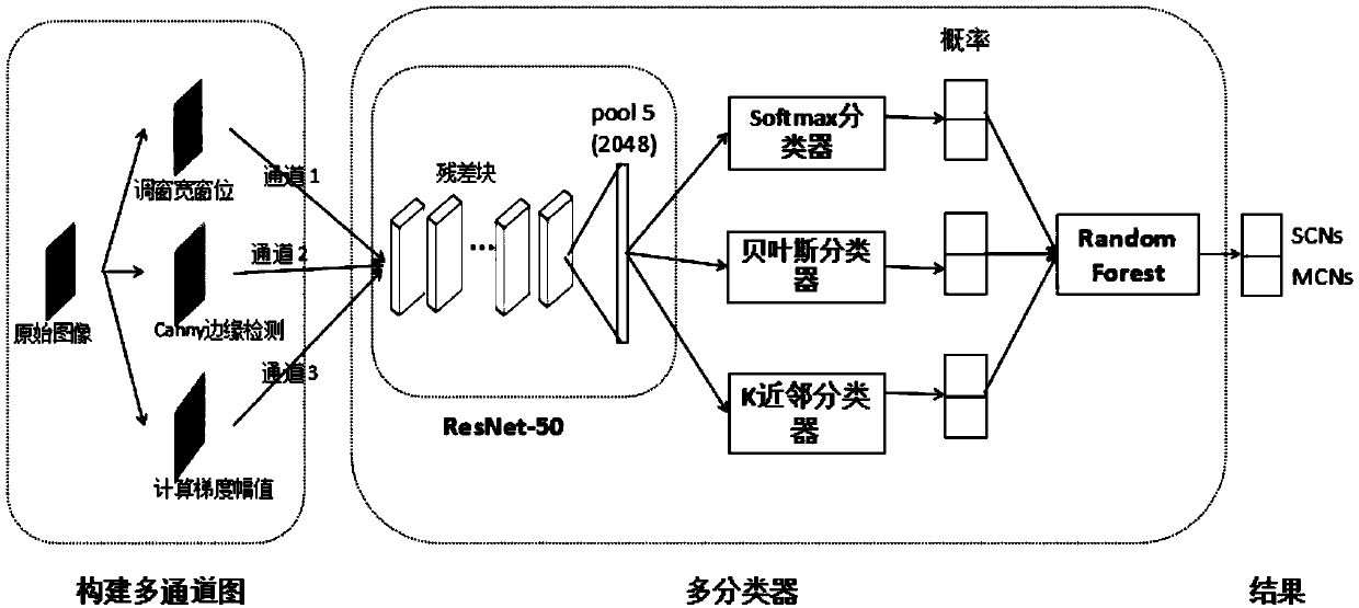
[0047] refer to Figure 1-Figure 4 , a method for classifying CT images of pancreatic cystic tumors based on multi-channel multi-classifiers, comprising the steps of:

[0048] 1) if figure 2 As shown in the multi-channel part of , the original image is adjusted for window width and level operation, Canny edge detection and gradient amplitude calculation are performed to enhance the edge features. The process is as follows: 1.1) Adjust the window width and level: by adjusting the appropriate window width and level To observe the cystic tumor of the pancreas to make it more clear. After adjustment, pancreatic cystic tumors are displayed in different simulated grayscales within a certain range. Above this range, pixel values ​​are displayed in full white. Conversely, pixel values ​​smaller than this range will be displayed in full black. The relationship between win...


PUM


Abstract

A pancreatic cystic tumor image classification method based on multi-channel multiple classifiers comprises the following steps: 1) performing window width and window level adjustment on an original image, and performing Canny edge detection and gradient amplitude calculation to enhance edge features; 2) adopting a ResNet to perform end-to-end training on a multi-channel graph, using an output ofa pool5 layer as extracted features, using a Bayesian classifier and a KNN classifier to perform classification, and obtaining the classification probabilities; and 3) using a random forest classifierto classify the obtained 3 different probabilities to get a final result, wherein a random forest is composed of multiple decision trees. The pancreatic cystic tumor image classification method basedon multi-channel multiple classifiers can automatically perform edge enhancement, and can improve classification accuracy.

Description

technical field [0001] The invention belongs to the technical field of image classification, and in particular relates to an image classification method. Background technique [0002] Usually, patients need to undergo a series of examinations before undergoing surgery, among which the observation of organs and lesions through computed tomography (CT) imaging is an indispensable method. Currently, physicians use their experience to determine whether cystic tumors of the pancreas are serous or mucinous. Existing image classification methods can be divided into traditional methods and deep learning methods according to their characteristics. Traditional methods first manually segment the tumor area; then, use different feature extractors to extract features, such as: texture features, structural features, etc., and then classify them with a classifier. The method of deep learning also needs to segment the original image first, then use the convolutional neural network to extr...

Claims


Application Information

Patent Timeline
IPC IPC(8): G06K9/62G06T7/00G06T7/13
CPCG06T7/0012G06T7/13G06T2207/10081G06T2207/30096G06V2201/03G06F18/24155G06F18/24323
Inventor 管秋胡海根李康杰陈峰黄志军王捷龚明杰姜娓娓陈胜勇
Owner ZHEJIANG UNIV OF TECH
Learn More