3D-printing femoral-condyle five-in-one integration osteotomy device and forming method thereof

A 3D printer and printing stock technology, applied in the field of medical devices, can solve the problems of loose prosthesis, many interference factors, and unequal osteotomy positions, and achieve the effect of avoiding displacement deviation, good surgical effect, and high surgical precision.

Pending Publication Date: 2018-11-13
THE SECOND AFFILIATED HOSPITAL ARMY MEDICAL UNIV +1
View PDF6 Cites 4 Cited by
  • Summary
  • Abstract
  • Description
  • Claims
  • Application Information

AI Technical Summary

Problems solved by technology

The two osteotomies of the femoral condyle require careful positioning and careful observation of the position of the osteotomy device. There are many interfering factors, and a slight deviation will cause the osteotomy position to be wrong and the position of the femoral prosthesis to be poor, causing loosening of the prosthesis in the later stage. , the operation failed

Method used


Image

  • 3D-printing femoral-condyle five-in-one integration osteotomy device and forming method thereof
  • 3D-printing femoral-condyle five-in-one integration osteotomy device and forming method thereof
  • 3D-printing femoral-condyle five-in-one integration osteotomy device and forming method thereof

Examples


Embodiment 1

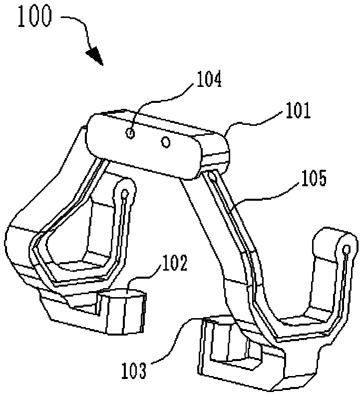
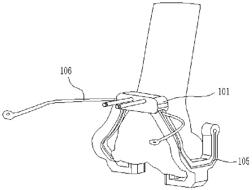
[0037] Such as Figure 1 to Figure 4 As shown, this embodiment provides a corresponding structural diagram of a 3D printed femoral condyle 5-in-1 integrated osteotomy device, a schematic diagram of the structure used, and a schematic diagram of the structure after use. Specifically, the 3D printed femoral condyle 5 provided in this embodiment The 1 integrated osteotomy device includes: a femoral osteotomy device model 100 adapted to the patient's affected femoral condyle, which is provided with a fitting surface 101 in front of the femoral condyle, a fitting surface 102 of the medial femoral condyle and a fitting surface 103 of the lateral femoral condyle , the anterior fitting surface 101 of the femoral condyle is used for fixing with the femoral condyle; the femoral osteotomy device model 100 has the characteristic that it is not easily deformed under force; the femoral osteotomy device model 100 is shaped by 3D printing.

[0038]It should be noted that during application, t...

Embodiment 2

[0047] The forming method of the 3D printed femoral condyle 5-in-1 integrated osteotomy device provided in this embodiment includes the following steps:

[0048] Step 1. Obtain patient data, use MRI to scan the patient's ankle joint to femoral head on the affected side, and save and output the scanned data in DICOM format; take X-rays of the whole lower limbs, and routinely take full-length X-rays of both lower limbs;

[0049] Step 2. Model reconstruction: process the scan data in step 1, import the DICOM data into MIMICS17.0 software for pre-processing, use the threshold value command to extract the knee joint tissue through the difference of MRI gray value, and generate 1:1 3D Lower limb bone model;

[0050] Step 3. Optimizing the 3D lower limb bone model in step 2 for preoperative simulated osteotomy;

[0051] Step 4. Preoperative simulation. Determine the landmark points of the model osteotomy in Materialize 3-matic and SIEMENS NX 9.0 software, determine the three-dimensi...


PUM


Abstract

The invention provides a 3D-printing femoral-condyle five-in-one integration osteotomy device and a forming method thereof, and relates to the technical field of medical apparatuses and instruments. For solving the technical problem that errors are easy to generate in existing total knee replacement femoral-condyle osteotomy, the 3D-printing femoral-condyle five-in-one integration osteotomy devicecomprises a femoral osteotomy device model matched with the affected-side femoral condyle of a patient, the femoral osteotomy device model is provided with a femoral-condyle front fitting face, a femoral-medial-condyle fitting face and a femoral-lateral-condyle fitting face, and the femoral-condyle front fitting face is used for fixed with the femoral condyle; the femoral osteotomy device model is subjected to stress and is not front to deformation, and is formed in a 3D-printing mode. Two osteotomy processes can be merged into one process, femoral-condyle osteotomy is completed one time, theusing quantity of osteotomy guiding plates is decreased, and deviations are reduced.

Description

technical field [0001] The invention relates to the technical field of medical devices, in particular to a 3D printed femoral condyle 5-in-1 integrated osteotomy device and a forming method thereof. Background technique [0002] Artificial knee replacement is the main treatment for advanced knee osteoarthritis and other diseases. During the operation, femoral condyle and tibial plateau osteotomy are required. The femoral condyle has 5 osteotomy surfaces, which are completed in 2 times by two osteotomy devices. The first is a horizontal plane osteotomy device, which cuts out the first osteotomy surface of the femoral condyle, and the second is a 4-in-1 osteotomy guide plate device, it cuts out two front and back faces of the femoral condyle on the first cut out horizontal plane. The two osteotomies of the femoral condyle require careful positioning and careful observation of the position of the osteotomy device. There are many interfering factors, and a slight deviation will...

Claims


Application Information

Patent Timeline
Patent Type & Authority Applications(China)
IPC IPC(8): A61B17/16
CPCA61B17/1657A61B17/1675
Inventor 王敏张佳思李伟浩代明君王小鹏柯松徐源周媛媛刘娇
Owner THE SECOND AFFILIATED HOSPITAL ARMY MEDICAL UNIV
Why Eureka
Try Eureka