
Magnetic resonance apparatus and method for creating maximum intensity projection

A technology of maximum intensity projection and magnetic resonance, which is applied in magnetic resonance measurement, measurement device, measurement using nuclear magnetic resonance imaging system, etc., which can solve the problems of high extra cost and unavailability of diagnosis.

Active Publication Date: 2018-10-09
SIEMENS HEALTHCARE GMBH
View PDF4 Cites 0 Cited by
  • Summary
  • Abstract
  • Description
  • Claims
  • Application Information

AI Technical Summary

Problems solved by technology

In the worst case, MR images produced according to the state of the art can be classified as diagnostically unusable, resulting in a 6 to 10 minute length of time to reacquire the data or even a rescheduled appointment with the patient
The resulting additional costs are correspondingly high

Method used


Image

  • Magnetic resonance apparatus and method for creating maximum intensity projection
  • Magnetic resonance apparatus and method for creating maximum intensity projection
  • Magnetic resonance apparatus and method for creating maximum intensity projection

Examples


Embodiment Construction

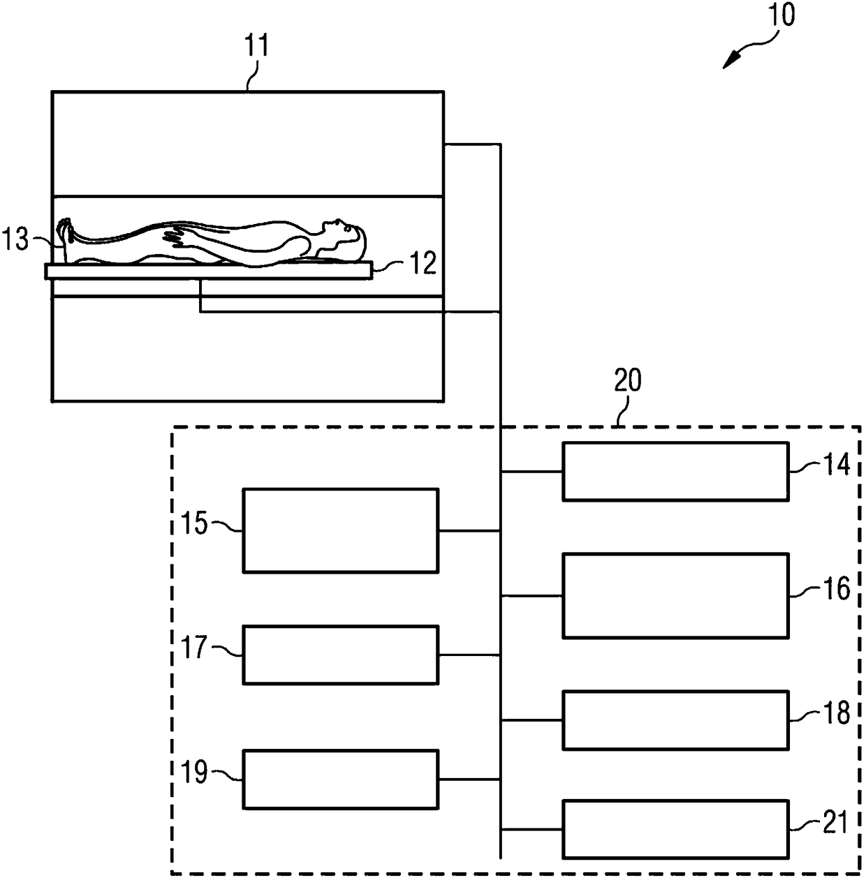
[0041] refer to figure 1 A magnetic resonance device is explained, with which MR data of the examination object 13 can be acquired, as explained below. The magnetic resonance apparatus 10 has a magnet 11 for generating a polarization field B0 , wherein a subject 13 positioned on a couch 12 enters the magnet 11 in order to record position-encoded magnetic resonance signals from the subject 13 there. Coils used for signal recording, such as whole body coils or local coils, are not shown for clarity. The invention can be applied to so-called parallel imaging, in which MR signals are recorded simultaneously with a plurality of local coils (coil array of local coils). By injecting a high-frequency pulse and switching on a magnetic field gradient, the magnetization generated by the polarization field B0 can be deflected from the equilibrium position and encoded locally, and the resulting magnetization detected by the receiver coil. How to generate MR images by radiating RF pulses ...


PUM


Abstract

In a method and magnetic resonance (MR) apparatus (10) for creating a maximum intensity projection of a volume of an examination subject (13), MR data are acquired for a number of slices (1) of the volume, and an MR image is reconstructed for each of the slices that MR data have acquired in order to create a maximum intensity projection for each of the slices (1). The maximum intensity projectionof the respective slice (1) is shown on a display (15). The slices (1), which have a thickness of at least 15 mm, have various slice directions, in order to display the maximum intensity projections from various directions.

Description

technical field [0001] The invention relates to a method and a magnetic resonance system for generating maximum intensity projections by means of a magnetic resonance system. Background technique [0002] Magnetic resonance cholangiopancreatography (MRCP) is a non-invasive method for visualization of the (fluid-filled) hepatobiliary system (the bile duct system and the main efferent duct of the pancreas) by means of magnetic resonance imaging. For this purpose, according to the prior art, high-resolution T2-weighted three-dimensional recordings are performed with extremely long echo times, in which maximum intensity projections are performed for diagnosis from different viewing angles (so-called radial viewing angles). [0003] Since a large amount of data cannot be collected under the breath hold state (Breath Hold), according to the prior art, data must be collected in a longer period of time (longer than the breath hold state), so that the patient must hold his breath man...

Claims


Application Information

Patent Timeline
Patent Type & Authority Applications(China)
IPC IPC(8): G01R33/56A61B5/055
CPCA61B5/055G01R33/56A61B2576/02G01R33/4835G01R33/546G01R33/5608G01R33/5635G01R33/56509G01R33/34A61B5/0044
Inventor D.保罗F.卡林奇W.霍杰
Owner SIEMENS HEALTHCARE GMBH
Learn More