
Method and associated apparatus for setting the contrast of a multi-energy CT image representation

A CT image and contrast technology, applied in the field of setting equipment, computed tomography system, and multi-energy CT imaging, can solve the problems of unsuitable radiology and large variability

Active Publication Date: 2022-08-09
SIEMENS HEALTHCARE GMBH
View PDF0 Cites 0 Cited by
  • Summary
  • Abstract
  • Description
  • Claims
  • Application Information

AI Technical Summary

Problems solved by technology

[0010] However, "unified filters" of the type described above are not suitable for radiology because of the greater variability of structures to be visualized (e.g. lesions) in radiology

Method used


Image

  • Method and associated apparatus for setting the contrast of a multi-energy CT image representation
  • Method and associated apparatus for setting the contrast of a multi-energy CT image representation
  • Method and associated apparatus for setting the contrast of a multi-energy CT image representation

Examples


Embodiment Construction

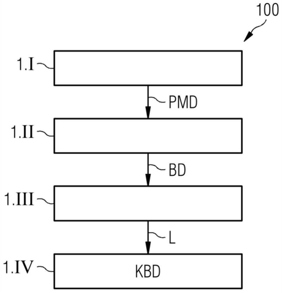
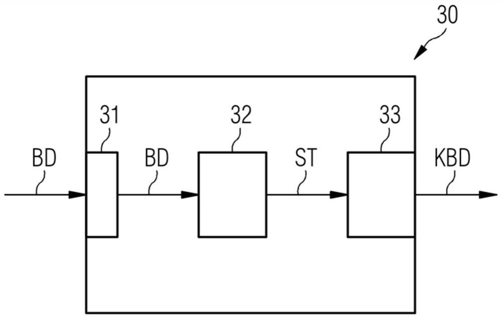
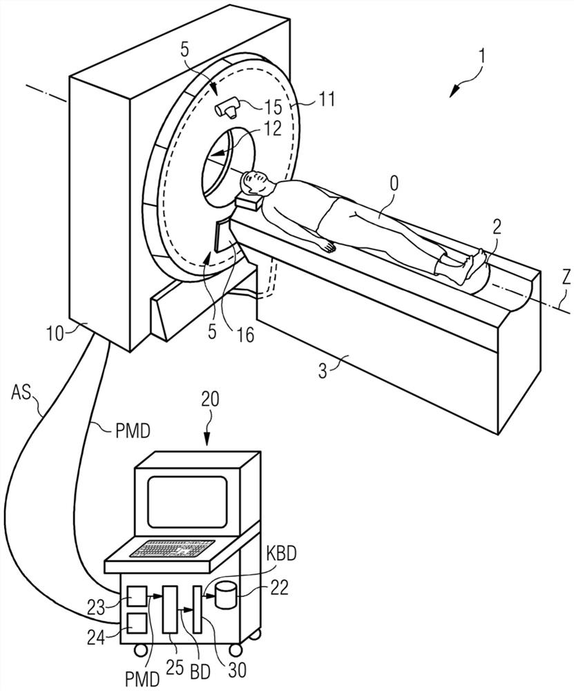
[0039] figure 1 The illustrated flowchart 100 illustrates a method for setting the contrast of a multi-energy CT image representation in accordance with an exemplary embodiment of the present invention. First, at step 1.1, a plurality of multispectral projection measurement data sets PMD of the examination area of ​​the patient are acquired. Next, at step 1.II, a plurality of multispectral image datasets BD are reconstructed based on the multispectral projection measurement dataset PMD. At step 1.III, the lesion L to be examined is identified in an automated manner based on the multispectral image dataset BD. Recognition can be achieved by means of a recognition step, which has been trained by means of a machine learning process to detect anomalies, in this case lesions L, by automated means. At step 1.IV, the multispectral image dataset is then weighted such that the contrast between the lesion L and its environment increases compared to uniform weighting. For example, suc...


PUM


Abstract

The present disclosure relates to contrast-enhanced reconstruction of spectral CT image data. A method for setting the contrast of multi-energy CT image representations is described. In the method, a plurality of multispectral image datasets (BD) of an examination region of a patient (O) are received. Furthermore, automatic identification of structures to be examined (ST) is achieved based on multispectral image datasets (BD), and automatic combination of differently weighted multispectral image datasets (BD) is achieved such that compared to uniform weighting, the The contrast between the inspected structure (ST) and its environment is improved. Furthermore, a multi-energy CT imaging method is described. An image setting device (30) is also described. Furthermore, a computed tomography system (1) is described.

Description

technical field [0001] The present invention relates to a method for setting the contrast of a multi-energy CT image representation. The invention also relates to a multi-energy CT imaging method. The invention also relates to a setting device. Furthermore, the present invention relates to a computed tomography system. Background technique [0002] Existing imaging techniques are often used to aid in the generation of 2D or 3D image data that can be used to visualize the imaged inspection object, among other applications. [0003] Imaging techniques are usually based on the detection of X-ray radiation, during which process primary data is generated, known as projection measurement data. For example, projection measurement data can be acquired by means of a computed tomography system (CT system). In a CT system, the combination of an X-ray source arranged on a gantry and an oppositely arranged X-ray detector usually rotates around a measurement chamber in which the objec...

Claims


Application Information

Patent Timeline
Patent Type & Authority Patents(China)
IPC IPC(8): A61B6/03
CPCA61B6/03A61B6/42A61B6/481A61B6/482A61B6/5205G06T5/007G06T2207/10081G06T5/50G06T5/009A61B6/5217A61B6/032G06T7/0012G06T2207/20208G06T2207/30096G06T2207/20081
Inventor M·里奇S·施密特
Owner SIEMENS HEALTHCARE GMBH
Learn More