
Method and apparatus for identifying pathological picture

一种图片、病理的技术,应用在医疗领域,能够解决耗时耗力、不同识别结论、准确率不太理想等问题,达到提高效率、提高准确率的效果

Active Publication Date: 2018-09-28
SUN YAT SEN UNIV CANCER CENT
View PDF7 Cites 23 Cited by
  • Summary
  • Abstract
  • Description
  • Claims
  • Application Information

AI Technical Summary

Problems solved by technology

A pathological picture has tens of thousands of pixels in length and width. To make an identification, a doctor with rich experience in nasopharyngeal carcinoma is generally required to observe carefully under the condition of 40 times magnification. The whole process Not only is it time-consuming and labor-intensive, it relies on experience, and the accuracy rate is not ideal, but doctors with different experience may have different identification conclusions for the same pathological picture.

Method used


Image

  • Method and apparatus for identifying pathological picture
  • Method and apparatus for identifying pathological picture
  • Method and apparatus for identifying pathological picture

Examples


Embodiment Construction

[0026] In order to make the purpose, technical solutions and advantages of the embodiments of the present invention more clear, the embodiments of the present invention will be further described in detail below in conjunction with the accompanying drawings. Here, the exemplary embodiments and descriptions of the present invention are used to explain the present invention, but not to limit the present invention.

[0027] The technical terms involved in the embodiments of the present invention are briefly described below.

[0028] Accuracy rate: Accuracy=(number of samples predicted correctly) / (total number of samples).

[0029] Precision: Precision=(number of samples predicted to be 1 and correctly predicted) / (number of samples predicted to be 1).

[0030] Recall rate: Recall = (the number of samples predicted to be 1 and correctly predicted) / (the number of samples whose true situation is 1).

[0031] Top5 error rate: imagenet images usually have 1,000 possible categories, ...


PUM


Abstract

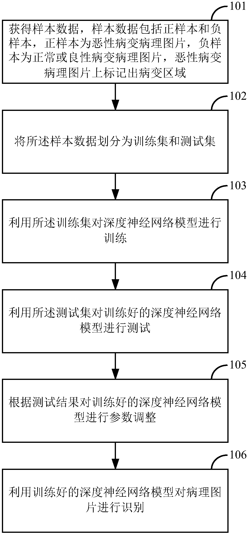
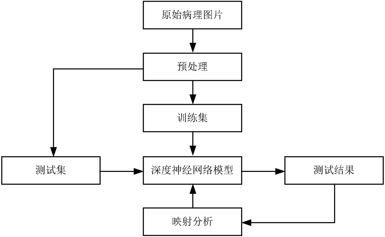
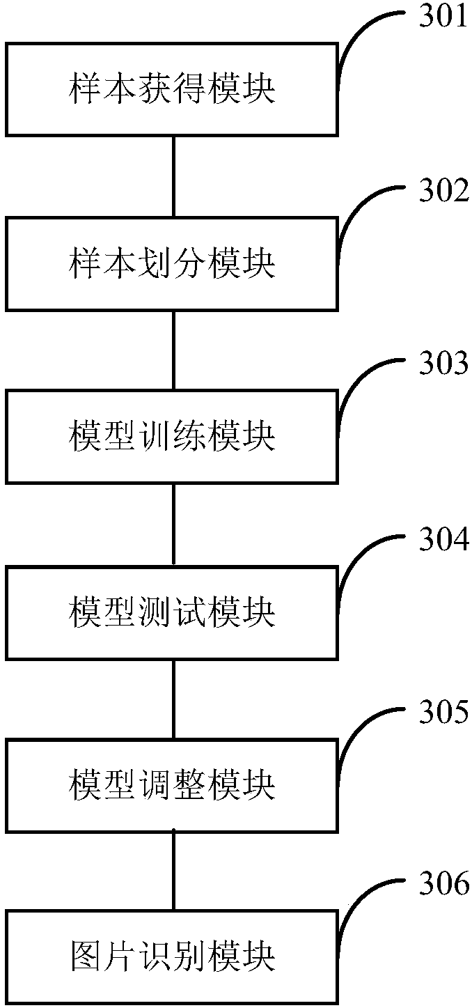
The present invention discloses a method and an apparatus for identifying a pathological picture. The method comprises: obtaining sample data, wherein the sample data comprises a positive sample and anegative sample, the positive sample is a malignant lesion pathological picture, the negative sample is a normal or a benign lesion pathological picture, and a lesion area is marked on the malignantlesion pathological picture; dividing the sample data into a training set and a test set; training a deep neural network model by using the training set; testing the trained deep neural network modelby using the test set; performing parameter adjustment on the trained deep neural network model according to a test result; and identifying the pathological picture by using the trained deep neural network model. According to the method and apparatus disclosed by the present invention, the efficiency and accuracy of pathological picture identification can be improved.

Description

technical field [0001] The invention relates to the field of medical technology, in particular to a method and device for identifying pathological pictures. Background technique [0002] At present, patients suspected of having nasopharyngeal carcinoma need to submit the pathological pictures of their nasopharynx to the corresponding doctor for observation, and identify the normal, benign and malignant pathological pictures. A pathological picture has tens of thousands of pixels in length and width. To make an identification, a doctor with rich experience in nasopharyngeal carcinoma is generally required to observe carefully under the condition of 40 times magnification. The whole process Not only is it time-consuming and labor-intensive, it relies on experience, and the accuracy rate is not ideal, but doctors with different experience may have different identification conclusions for the same pathological picture. Contents of the invention [0003] An embodiment of the p...

Claims


Application Information

Patent Timeline
Patent Type & Authority Applications(China)
IPC IPC(8): G06T7/00G06N3/04
CPCG06T7/0012G06T2207/20081G06T2207/30096G06N3/045G06T7/0014G06T2207/20084G06T2207/30024G16H50/70G16H50/20G16H30/40G06N3/08G06T7/0016G06F18/2148
Inventor 云径平王智
Owner SUN YAT SEN UNIV CANCER CENT
Learn More